|
Integrating Imaging in Diagnosing
Equine Lameness
Tracy A. Turner, DVM, MS; Dipl.ACVS,
St. Paul, Minnesota 55108
There are several different methods of imaging the equine digit.
Imaging is of utmost importance because it will provide pathologic
and physiologic information necessary to treat the specific
condition. Imaging can be divided into anatomic and physiologic
imaging methods. Anatomic imaging modalities include radiology,
ultrasonography, computer-aided tomography, and magnetic resonance
imaging. Physiologic imaging modalities include scintigraphy and
thermography.
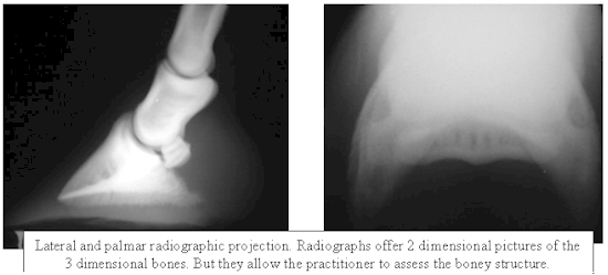
Radiologic techniques are the most commonly used to evaluate the
horse for lameness. Utilizing plain film radiography, it requires
multiple projections to evaluate any area. Essentially the
practitioner must attempt to draw conclusions about a three
dimensional object utilizing 2 dimensional pictures. Occassionally
it becomes necessary to utilize radiographic techniques that
provide more information. Contrast radiography is one such
technique that provides information amount the articular cartilage
and surfaces. It is of particular value in determining whether
subchondral cysts commmunicate with the joint or in dilineating a
subcutaneous tract. Generally, 5 to 10 ml of contrast injected
into the joint is adequate. Pathologic diagnoses are usually made
by radiography in conjunction with clinical examination.
Ultrasonographic examination can be used to assess any soft tissue
in the horses body. The deeper the tissue that needs to be
evaluated the lower wavelength probe needs to be used. The tissues
are examined for changes in echogenicity. Changes in echogenicity
correspond to changes in the tissue. Ultraonography is most useful
in the evaluation of tendons and ligaments but it also can be used
to evaluate muscle and cartilage.
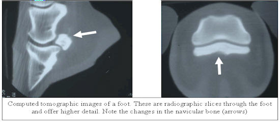
Magnetic resonance imaging and computer-aided tomography are both
interesting and high detail anatomic imaging tools. However, at
this time they are of little value to the practicing veterinarian
but may help provide insight to researchers about the pathology.
Physiologic imaging techniques would be those techniques that
provide the evaluator with an image that reflects physiologic
processes. Unlike anatomical imaging that reflects structure,
these images give insight into metabolism or circulation.
Thermography and scintigraphy provide the examiner with the
opportunity to examine the entire horse. When combined with a
thorough clinical examination, these methods are extremely useful
in identifying injuries that may have otherwise gone undetected.
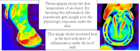
Thermography is the pictorial representation of the surface
temperature of an object. It is a non-invasive technique that
measures emitted heat. A medical thermogram represents the surface
temperatures of skin making thermography useful for the detection
of inflammation. This ability to non-invasively assess
inflammatory change, makes thermography an ideal imaging tool to
aid in the diagnosis of certain lameness conditions in the horse.
The circulatory pattern and the relative blood flow dictate the
thermal pattern which is the basis for thermographic
interpretation. The normal thermal pattern of any area can be
predicted on the basis of its vascularity and surface contour.
Skin overlying muscle is also subject to temperature increase
during muscle activity. Injured or diseased tissues will
invariably have an altered circulation. One of the cardinal signs
of inflammation is heat which is due to increased circulation.
Thermographically, the "hot spot" associated with the localized
inflammation will generally be seen in the skin directly overlying
the injury. However, diseased tissues may in fact have a reduced
blood supply either due to swelling, thrombosis of vessels, or
infarction of tissues. With such lesions the area of decreased
heat is usually surrounded by increased thermal emissions,
probably due to shunting of blood.
Scintigraphy utilizes polyphosphonate radiopharmaceuticals
administered by intravenous injection and followed by measurement
of the distribution of the pharmaceutical by a gamma camera.
Concentrations of the pharmaceutical can be detected, as the
polyphosphonates bind rapidly to exposed hydroxyapatite crystal.
This is generally in areas where bone is actively remodelling.
This is the basis of the bone scan but prior to this the
distribution of the drug goes through two other phases. It is
these phases that can be useful to evaluate soft tissue changes.
There are three phases, the vascular phase, the soft tissue phase,
and the bone phase. The vascular phase or blood pool phase begins
immediately after injection of the pharmaceutical. This phase is
dependent on local variations in vascular supply. The most common
clinical application for vascular phase scintigraphy is
determination of patency of blood vessels. The second phase or
soft tissue phase scintigraphy is performed while most of the
pharmaceutical is in the extracellular fluid (ECF). This usually
begins 1-2 minutes post pharmaceutical injection and lasts until
significant uptake of the polyphosphanate by bone, usually 1-2
hours. The distribution of the radiopharmaceutical during this
phase is due to local blood flow, capillary density, capillary
permeability and regional ECF volume. Because inflammation causes
an increase in blood flow, capillary permeability and ECF volume,
inflamed tissues accumulate high levels of radiopharmaceutical.
This is the basic principle behind evaluation of soft tissue
injuries by scintigraphy. The bone phase is the most useful in
that the uptake of the radiopharmaceutical always increases around
areas of increased remodelling or vascularity. Since injured bone
is under going more rapid remodelling, this is the basis for using
bone phase to detect injuries. Scintigraphy has been most useful
for the detection of lesions in bone and ligaments. Scintigraphy
has been particularly useful in the identification of enthesopathy
(damage to the insertions of tendons and ligaments on bone).
The purpose of any lameness examination is to be able narrow the
problem to a regional diagnosis. Once a regional diagnosis has
been made it is possible to assess the area utilizing some type of
anatomical imaging modality. Assessment of those anatomical
changes serves as the basis for any pathologic diagnosis that may
be made, as well as, being important in determining prognosis. For
these purposes radiography and ultrasonography are complimentary.
Radiography provides information regarding the boney tissues.
Radiographs reflect change that has happened. Ultrasonography
provides information about boney comntour but moreimportantly
provides insight to the soft tiisues that connect bone or provide
support. Sonography can give much better insight into the activity
of a lesion. That is, is the lesion active or not, do the soft
tissues changes reflect an ongoing process or is it a chronic
process. In addition, sonography can provide information about
joint capsule, collateral ligaments, the consisitncy of joint
fluid, and provide insight into the articular cartilage.
However, the pursuit of a regional diagnosis can be difficult.
There are 3 instances where this can be frustrating. One, when
diagnostic analgesia has failed to eliminate the lameness; two,
when the lameness is too subtle to avail itself to diagnostic
analgesic techniques, and three, when the patient is not amenable
to handling or injection. In these cases, other methods must be
used to evaluate the patient. This is where physiologic imaging
modalities can be so useful. By providing insight into physiologic
changes in the tissues, this can lead the examiner to evaluate
those areas utilizing anatomic imaging methods.
Another area in lameness evaluation where imaging can be useful is
in preventing injury. This requires the early detection of the
physiologic change of injury. Although, the frequent use of an
anatomical imaging modality can discover change in one region,
physiologic imaging allows the assessment of the entire animal on
a routine basis.
The utlization of imaging modalities in the diagnosis and
treatment of equine lameness is absolutely necessary. These are
the only reliable methods to assess the type and severity of the
injury. In addition, the routine use of any method can provide
insight into the stresses and strains of the athlete.

|