Low Heels In The Hind Feet - An Often Overlooked Problem
A look at two treatments with succesful clinical results
Reprinted with permission of the American Farriers Journal.
Original printed in the 2007 March / April issue of the American Farriers Journal
Stephen E. O'Grady DVM and J.G. Merriam DVM
1. Introduction
Low, underrun or collapsed heels
affecting the health of the foot or
as a cause of lameness has been
well documented in the front limbs.1
However, very little information has been
written concerning the effects of low or
damaged heels in the hind limbs.
Horses with structural damage to the
heels of the hind feet will suffer the same
consequences associated with the hoof
capsule as noted in the front feet, but the
hind feet don't appear to be affected with
disease of the internal structures as noted
in the forefeet.
This difference may be due to the
anatomy of the hind limbs and the
propulsionary function of the hind feet.
Damage to the structures of the hind feet
may be well advanced before lameness
is noted. Underrun or collapsed heels in
the hind feet may lead to a subtle bilateral
lameness, which is often attributed
to hock, stifle or back pain.
Lameness issues in the hind limbs are
often localized to the proximal suspensory
ligament, the hocks or the stifle.
Part of the therapy for lameness
involving these structures is to raise the
heels of the hind feet regardless of the
conformation of the hind foot. Long egg
bar shoes or egg bar shoes with wedge
pads are generally used for this purpose.
Yet there is absolutely no documentation
that confirms that heel elevation
exerts significant influence on any part of
the hind limb anatomy above the distal
interphalangeal (DIP) joint. 2
Furthermore, heel elevation applied to
the hind feet that have existing low heels
or underrun heels appears to damage the
heels further, leading to an additional
lameness problem in and of itself. The
lameness caused by damage to the heels
is often diagnosed secondary to the
affected ligament or joint for which the
heels were originally elevated.
 |
2. Clinical Examination Of The foot
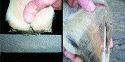
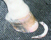
Abnormal heel conformation of the
hind feet is easy to recognize. When
looking at the limb from the side, the
digit will show a broken back hoofpastern
axis. The slope of the coronary
band from the toe to the heel will have an
acute angle. The bulbs of the heels will
have a bending appearance and can be
seen lying against the shoe palmar to the
end of the heel. The dorsal hoof wall
begins to take on a "bull nosed" appearance
(Figure 1A).
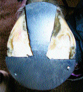
Looking at the foot from behind, the
frog is situated well below the hoof wall
and the frog can be seen to prolapse
down between the two branches of the
shoe (Figure 1B).
The frog is generally large from the
constant stimulation with the ground.
The clinical appearance of a hind foot
with the heels damaged by an egg bar
shoe and a wedge pad are much the same (Figures 2A and 2B). The broken back
hoof pastern axis will not be as marked
and the angle of the coronet will not be
as acute, but the damage to the heels and
soft tissue structures heel of the foot will
be greater due to the continuous pressure
exerted by the length of the shoe and the
wedge pad.
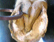
Upon removing the shoe, the end of
the heel of the hoof wall is located well
forward from the base of the frog. The
horn tubules will be parallel with the
ground. The hoof wall at the heel will be
thin, there will be no angle to the sole and
the bars will be absent. The whole frog
will be pushed down below the hoof wall
(Figure 3).
When the foot is placed on the
ground, total weight bearing will be
placed on the frog and many horses are
reluctant to stand on it when the opposing
limb is lifted off the ground.
Viewing the ground surface of the
foot, there will be a "trough" noted
between the apex of the frog and the
inner branch of the shoe at the toe. Hoof
testers placed on either side of the heel at
the angle of the sole will elicit a painful
response (Figure 4).
|
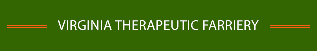
| Fig 1A. Note the arrow denoting the end of the heel |
 |
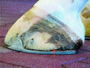
| Fig 1B. Note the position of the frog below the hoof wall at the heels. |
 |
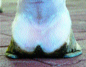
| Fig 2A. Again note the arrow at the end of the heel. |
 |
 |
| Fig 2B. Note how pressure is placed on the soft tissue structures of the heel by the wedge pad and the egg bar shoe. |
Fig 3.The left photo shows damage to the heel with an open shoe. The photo on the right shows where the base of the frog was weight bearing on the egg bar shoe. |
 |
 |
| Fig 4. Pressure from hoof testers will also show movement due to separation in the damaged heels. |
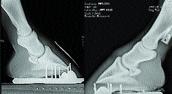
Fig 5. Note the position of P2 relative to P3 in both radiographs.This places the load on the palmar section of the foot. |
3. Radiographs
A lateral radiograph of the hind foot
will show a broken back hoof pastern
with the second phalanx (P2) being
pushed palmarly and distally relative to
the distal phalanx (P3) during weight
bearing (Figure 5). This places excessive
stresses on the palmar section of the
joint capsule.
The palmar margin (palmar angle) of
the distal phalanx is lower when
compared with the dorsal margin of the
distal phalanx. Damage to the heels of
the hoof capsule can be noted below the
palmar process of the distal phalanx as
lucent areas in the hoof capsule.
The sole depth below the dorsal
margin of P3 is markedly increased relative
to the heel and the perimeter of the
distal phalanx can be seen migrating
toward the dorsal hoof wall. This is what
causes the "bull nose" appearance of the
dorsal hoof wall. The soft tissue structures
in the palmar section are noted to be
lying against the shoe.
 |
| Fig 6A. A foot that was shod with an egg bar shoe. Note the damage to the heels. |
 |
| Fig 6B.The same foot in Figure 6A after rasping the heels down to solid tissue and leaving the horse barefoot for 6 weeks. |
 |
| Fig 7. Excess hoof wall is removed from toe quarter to toe quarter. |
 |
| Fig 8. A 2-degree wedge pad cut out so weight bearing is concentrated over the frog. |
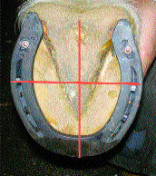 |
| Fig 9. Shoe fitted to hind foot after frog is displaced so it is on the same plane as the heels. Note a line drawn across the widest part of the foot lies in the middle of the shoe. Note breakover has been created in the shoe with a grinder. |
4. Treatment
Damage to the heels
of the hind feet is often
easier to improve than
damage to the forefeet,
possibly due to the
difference of the load
encountered on the
hind limbs vs. the fore
limbs. Two methods
can be employed to
treat this condition.
First, allowing a horse to go without hind shoes - if possible - for 4 to 8 weeks can be very effective.
This approach can also be used with
horses that are resting due to proximal
suspensory ligament disease. The shoes
are removed and the hoof wall at the
heels is moved palmarly until solid structures
of the hoof wall are encountered.
The hoof wall at the toe is lowered appropriately
and the edges are rounded.
Over the next few weeks, the pressure
on the frog will compress and displace
the frog until it assumes the same plane
as the heels on either side (Figures 6A
and 6B).
If the horse needs to continue in work
and wear shoes, the approach will be
different. The shoes are removed and the
heels are moved palmarly until solid horn
is established. Excess dorsal hoof wall is
removed from toe quarter to toe quarter
(Figure 7). The prolapsed frog needs to
be compressed in order to have a flat,
even plane that includes both the heels
and the frog. The back section of a degree
pad is cut out to fit over the frog as a
mirror image. A thin strip extending
across the toe is left attached to the frog
wedge and two 4.5 race nails are placed
through this strip into the hoof wall at the
toe quarters to hold the frog wedge
directly over the frog (Figure 8).
An Animalintex self-contained poultice
is saturated with water and applied so it
envelops the whole foot. It is secured to the foot with brown
gauze and elastic tape. The horse is now
placed in a stall with a firm surface for 24
to 48 hours. During this time, the feet are
submerged in a bucket of water a few
times to keep the poultice saturated.
At the onset of applying the frog
wedge, the horse is given 2 grams of
phenylbutazone (Bute), as some horses
will show mild discomfort and develop
a digital pulse. Therefore, when
medication is suggested and used, both
authors contend that veterinary assistance
should be solicited when
performing this procedure.
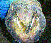
When the poultice is removed, the
frog will be compressed between the
heels forming a flat even surface that
includes the frog and both heels. The
horse can be shod immediately, or can be
placed in a stall bedded with sawdust for
an additional day to let the feet dry out.
The frog will be soft and can be
shaped further. Any additional horn at the
heels can be removed so the heels of the
hoof wall are solid and approach the base
of the frog - being careful to keep the
frog and both heels in the same plane. A
shoe can now be fitted and applied.
We fit shoes on the hind feet the same
as the front where a line is drawn across
the widest part of the foot and the shoe
is fitted so the line is placed in the middle
of the shoe.
In the hind feet, the branches of the
shoe may extend marginally beyond the
end of the heels (Figure 9). If additional
heel elevation is necessary, a wedge pad
or a bar wedge can be placed under the
heels as long as the shoe is fitted in the
manner just described. This will concentrate
the load under the frog and heels
rather than behind the heels, which is
the case with a long shoe.
5. Conclusions
The authors have used the frog pressure
and soaking technique on 15 horses
with low heels and prolapsed frogs. The
results have been excellent in all cases.
Damage to the heels of the hind feet
are much easier to resolve or improve
than the fore feet. This could be due to
the anatomy of the hind limb along with
the shape and function of the hind feet.
Once the frog has been repositioned and
the heel structures have grown, attention
to the foot prep is necessary to keep the
frog and heels in the same plane. The size
and placement of the shoe are equally
important in maintaining the health of the
heels of the hind feet.
References
- O'Grady, S. E. Strategies For Shoeing The Horse With Palmar Foot Pain, in Proceedings of 52nd Annual Convention Am Assoc Equine Practr, 2006: 209-217.
- Bushe T, Turner TA, Poulos PW, Harwell NM: The Effect Of Hoof Angle On Coffin, Pastern, And Fetlock Joint Angle, Proceedings of 33rd Annual Convention of Am Assoc of Equine Practnr, 1987: 689-699.
Dr. Steve O'Grady is a veterinarian
and a farrier. He operates Northern
Virginia Equine, a podiatry practice
located in Marshall, Va. Dr. Jay
Merriam is a partner in the
Massachusetts Equine Clinic, a full
service sport horse practice located in
Uxbridge, Mass.
|