White line disease: A review (1998–2018)Reprinted with permission from Equine Veterinary Education (EVE). |
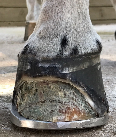 |
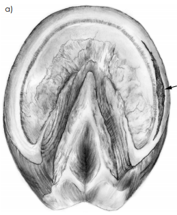
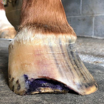
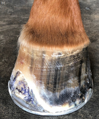
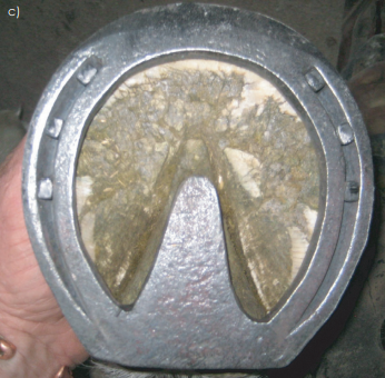
| Fig 1: Classic example of white line disease with separation atjunction ofstratum mediumandstratum internum. |
The destruction that occurs within the separation as a consequence of WLD remains superficial to the stratum internum and does not invade the dermis. White line disease always occurs secondary to a hoof wall separation (Turner 1998; O’Grady 2002; Moyer 2003; O’Grady 2006; Pleasant and O’Grady 2008; O’Grady 2011; Redding and O’Grady 2012). The disease has been termed seedy toe, hoof wall disease, hollow hoof, yeast infection, Candida and Onychomycosis. Seedy toe is a focal separation that occurs in the centre toe dorsal to the crena marginalis of the distal phalanx. The separation may extend dorsally in the hoof wall directly above the origin but does not deviate laterally or medially. This hoof wall defect appears to be associated with the crena marginalis (T. D. Burns, unpublished data 2016).
Onychomycosis denotes a mycotic disease that originates in the nail bed of the human and the dog. By contrast, in WLD the infection originates at the solar surface of the hoof and migrates proximally, approaching the coronet but never invading this structure. Keratinophilic fungi are often isolated from separated areas of the hoof wall (Kuwano 1998; Turner 1998; Ball 2000); however, in many cases of WLD, the pathogens cultured initially are purely bacterial or a mixed population of bacterial and fungal organisms (Turner 1998; O’Grady 2006). Therefore, onychomycosis may not be the appropriate term when referring to white line disease in the horse (O’Grady 2006). Furthermore, since the term ‘white line disease’ lacks a definitive definition, perhaps it should be considered a syndrome leading to a disease. The syndrome being symptoms such as a hoof wall separation, combined with a hoof capsule distortion leading to disease which is a pathophysiological response. Finally, the term white line disease that is commonly used to describe this hoof issue is a misnomer, as the disease process does not affect the white line or zona alba’ of the horse’s hoof capsule (Fig 1).
 |
 |
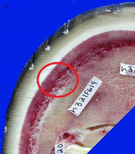 |
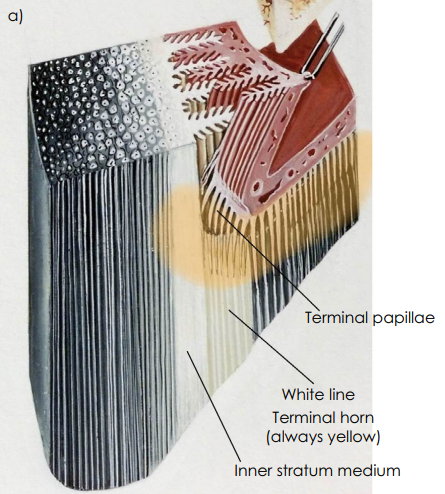
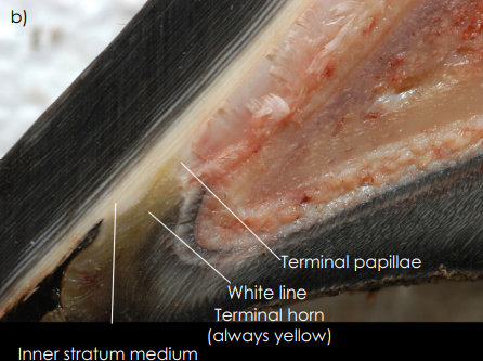
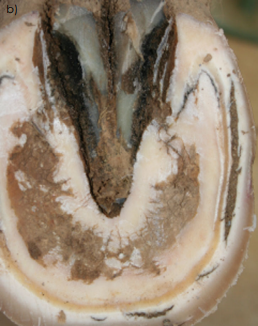
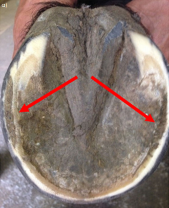
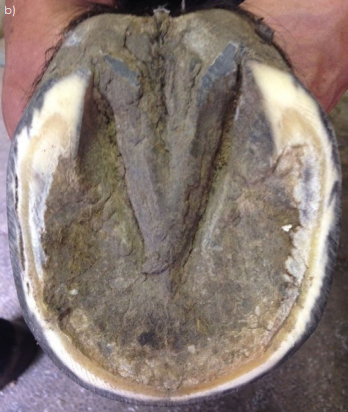
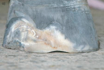
| Fig 2: Arrangement of the anatomical structures on the distal hoof. Orange highlights the growth zones in the distal hoof in (a). (b) shows separation present in the Inner section of the stratum medium that is nonpigmented and the stratum internum and (c) shows the striations (red circle) in the white line (Zona Alba) (a and b Courtesy of Dr Chris Pollitt; c Courtesy of Dr Jeff Thomason). |
Anatomy of the hoof wall
A closer look at the hoof wall is necessary to appreciate the anatomical location where WLD occurs.
The hoof wall consists of three layers:
The stratum externum arises from the perioplic epidermis and forms the thin outer layer of keratinised cells that give the wall its smooth, glossy appearance. The stratum medium, arising from the coronary epidermis, forms the bulk of the hoof wall and is the densest part of the horny wall. It consists of cornified epidermal cells arranged in parallel horny tubules surrounded by intertubular horn, which grow distally from the coronary groove to the basal border. In all hooves the stratum medium is always nonpigmented (white) in the deepest inner layer. The stratum internum arises from the lamellar epidermis, is nonpigmented, and when combined with the dermal lamellae, is responsible for attaching the hoof wall to the distal phalanx. The distal end of each dermal lamellae is a set of papillae known as the terminal papillae. Proliferation of basal cells from the keratinised epidermal lamellae and tubular horn formed from the epidermis overlying the terminal papillae form the terminal horn that fills the space between the inner layer of the wall (stratum internum) and the sole as they grow distally towards the ground surface (Bragulla et al. 1998; Pollitt 2010). This structure forms the bond between the hoof wall and the sole known as the white line or zona alba. When observed from the solar surface, this white line or zona albicans is always yellow in colour, resembles frog horn with a striated appearance and has a plastic consistency when compared to the adjacent dorsal hoof wall or horny sole (Fig 2a–c).
Aetiology
The aetiology of WLD remains elusive and is currently undetermined. The problem has been diagnosed in horses worldwide. The incidence of WLD in the United States is unknown and would be hard to determine without a working definition; however, our ability to recognise and treat this hoof issue appears to be increasing. A recent study in a small European country showed the incidence of WLD to be 17.8%; however, the authors did not state how they arrived at this diagnosis (Holzhauer et al. 2017). White line disease can affect a horse of any age, sex or breed. One or multiple hooves may be involved, and the affected hooves can be barefoot or shod. One or multiple horses on the same farm may be affected. It is generally agreed that WLD is a multifactorial condition that develops secondary to an initial hoof wall separation or hoof wall defect (Turner 1998; O’Grady 2002; Moyer 2003; O’Grady 2006; Pleasant and O’Grady 2008; O’Grady 2011; Redding and O’Grady 2012).
The presence of hoof wall separations encountered during the practice of farriery could be considered routine. These separations can be located in the toe, quarter or heel or any combination of the above. They generally vary in depth from 0.5 to 2 cm and when these separations are explored with a probe; the clinician will readily reach solid hoof wall. The unanswered question is why, in a small percentage of cases, a small insignificant hoof wall separation will progress proximally under the hoof wall to become a large significant clinical entity. There is a large population of horses that have hoof wall separations yet do not develop WLD; therefore, there may be some type of genetic propensity or cellmediated immunity responsible for horses to develop this condition. A recent study correlated a genetic variant associated with a hoof wall separation in a certain breed of ponies (Finno et al. 2015).
Multiple other causes for WLD have been proposed, but none have been scientifically proven. Moisture may play a role as WLD is seen more in wet humid areas but it is also seen in hot arid conditions. Excessive moisture over time may soften the foot, allowing a separation to progress or allowing easier entry of dirt and debris into an existing separation. Continual bathing of competition horses, especially during the warmer months, appears to soften the horn and may contribute to the incidence of WLD in this population of horses. Excessively dry hooves, on the other hand, may form cracks or separations in the inner hoof wall, allowing pathogens to invade the horn.
If hoof wall separations are left unattended, dirt and debris become packed into the hoof wall defect and often result in progressive mechanical separation. Keratinopathogenic bacteria and fungi are commonly isolated from hoof wall separations of horses with WLD, particularly those with more extensive lesions (Turner 1998; O’Grady 2011; Redding and O’Grady 2012). However, it is generally believed that these microorganisms are opportunistic, secondary invaders found in the environment that enter the hoof wall through an existing separation or hoof wall defect and then exacerbate the hoof wall separation by the production of proteases that degrade keratin. The fact that WLD occurs in the avascular hoof wall and can be resolved with debridement alone further detracts from this being a predominately infectious process or the primary cause (Turner 1998; O’Grady 2002; Moyer 2003; O’Grady 2006; Pleasant and O’Grady 2008; O’Grady 2011; Redding and O’Grady 2012).
Mechanical stress or increased biomechanical forces placed on the inner hoof wall of horses with less than ideal hoof conformation appears to encourage a separation. The horn tubules of the stratum medium are arranged in a pattern of density going from a high density at the outer hoof wall to a low density at the inner hoof wall. The gradient of tubular density mirrors the moisture across the hoof wall and appears to be responsible for the smooth energy transfer across the hoof wall from the force of impact with the ground to the bone (Thomason et al. 1992; Pollitt 2010). As a result of the low tubular density, the inner section of the stratum medium is less rigid, softer and susceptible to compromise or failure. Therefore, it would seem logical that excessive force or stress on this part of the hoof wall created by leverage, or the various hoof capsule distortions would play a major role in the formation of hoof wall separations. Abnormal hoof conformation such as long toe and/or low heel, clubfoot (flexural deformities) and sheared heels are commonly seen in cases of WLD (O’Grady 2018).
Clinical signs
White line disease offers minimal threat to the soundness of the horse until damage to the attachment between the external lamellae, and the inner hoof wall is sufficient to allow movement and/ or a change in position of the distal phalanx within the hoof capsule. Most cases of WLD are first identified by the farrier during routine hoof care. In the early stages of WLD, a hoof wall separation is noted somewhere along the junction of the hoof wall and the sole (Fig 3).
  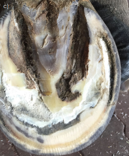 |
| Fig 3: Illustration in (a) shows a separation (arrow) in the inner hoof wall. (b) shows a separation in the hoof wall with the white line intact and (c) shows a separation that is more advanced |
 |
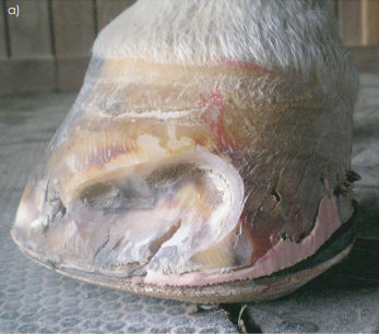
| Fig 4: Full-thickness hoof cracks in the distal one-third of the hoof capsule. Not the degenerating horn of the inner hoof wall that can be observed within the defect. Also, note the excessive hoof wall flares present on either side of the cracks |
The area of wall separation can vary in length and is typically filled with foreign material. Exploration of the separated area with a small thin loop knife or a probe reveals an undermined area of hoof wall of varying proportions. In the early stages of the disease, the foreign debris can be removed from the separated horn down to a solid connection between the inner hoof wall and the white line. This area may remain localised or it may progress to involve a larger area of the hoof wall. If the extent of the separation goes deeper, it will classically be filled with a grey-white powdery horn material. Other early warning signs of WLD, depending on the extent of the separation, may be tender soles as noted with hoof testers, occasional heat in the feet, and the sole may become increasingly flatter in the affected area. The hoof wall overlying the defect usually has a normal appearance, but the hoof capsule may have a flare or bulge outward in horses with an extensive separation. Furthermore, there may be some full-thickness hoof cracks originating at the distal border of the hoof capsule and extending proximally which encroach on the inner hoof wall (Fig 4).
 |
 |
| Fig 5: Hoof capsule distortion as a result of WLD. Extensive separation causing the distal phalanx to shift towards the side where the wall is undermined. Picture on the right shows same foot after trimming and the extent of the undermined horn. |
There may be slow hoof wall growth, poor consistency of hoof wall and a hollow sound will be detected when the hoof wall overlying the defect is percussed with a hammer (O’Grady 2006; O’Grady 2011; Redding and O’Grady 2012). Often the disease goes undetected unless there is a change in hoof wall conformation, or the horse begins to show discomfort. When the separation becomes more extensive and involves the toe and a quarter, a concavity (‘dish’) may be seen forming along one side of the hoof and a bulge will be present on the contralateral side directly above the affected area above the coronary band (Fig 5).
To explain the mechanism of this distortion – the distal phalanx is suspended within the hoof capsule by the circumferential lamellae when in the state of equilibrium. When a substantial separation affecting the epidermal lamellae attachment is present in the toe and either the medial or lateral quarter, the equilibrium is disrupted and the distal phalanx will shift towards the side of the foot with the separation, causing a concavity in the hoof wall on the contralateral side of the foot side of the foot.
Diagnosis
Lameness or hoof capsule abnormalities may not beobserved in the early stages of the disease. Hoof testerexamination does not always elicit a response. On the solar surface of the hoof, the sole/wall junction (white line) will bewider, softer and have a waxy texture. Exploring the innerhoof wall, which lies dorsal to the sole/wall junction, willgenerally reveal a separationfilled with white/grey powderyhorn material or there may be a black serous drainage fromthe separation. Further exploration with a blunt probe willreveal the depth and extent of the cavitation. The extent ofthe separation can be outlined by tapping the outer hoofwall with a hammer and then confirmed with radiographs. Ifthe separation is extensive, hoof testers applied over this areawill move the sole relative to the detached hoof wall leadingto a‘pinching’effect in the tissue located at the proximalextent of the defect. The movement of the sole against thewall will generate a painful response. If lameness is present, athorough lameness examination should be performedincluding local diagnostic analgesia to localise and confirmthe suspected area of discomfort and then followed by radiographs.
Radiographs
Radiography can be very informative and should be considered essential if the separation is extensive. Good quality radiographs show the extent of the hoof wall separation, and whether the distal phalanx has displaced within the hoof capsule. A podiatry study consisting of a lateral and horizontal dorsopalmar 0-degree views along with any lateral oblique views necessary to visualise the extent of the separation should be acquired to evaluate the foot (Fig 6).
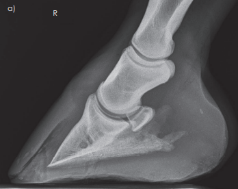 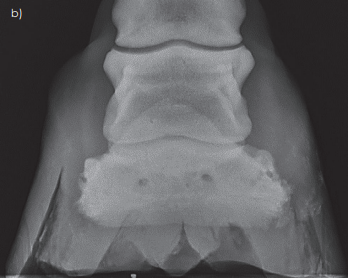 |
| Fig 6: Lateral and horizontal 0-degree DP radiographic views that show marked separations within the hoof wall. |
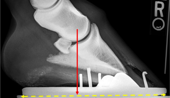 |
| Fig 7: Radiograph shows a marked separation in the dorsal hoof wall, displacement of the distal phalanx and a mild flexural deformity. Red arrow is the centre of rotation COR and the yellow dotted line is the proportions of the solar surface on either side of the COR. These guidelines can be used to decrease the leverage on the dorsal foot |
Laboratory
Laboratory findings have been unrewarding with regards to managing this disease.
Cultures may be of little value since the samples taken from the separations are contaminated with dirt and opportunistic organisms. Aerobic cultures usually reveal a mixed bacteria flora while anaerobic cultures are negative. Fungal cultures require a special media and time. The most common fungal species cultured are Pseudallescheria, Scopulariopsis and Aspergillus (Turner 1998; O’Grady 2006). A biopsy taken at the juncture between the normal and affected hoof wall shows a mixed population of microorganisms. These will generally include coccobacilli, yeast organisms and fungal spores. Inflammation in the laminar dermis will be seen deep to the affected area (Turner 1998).
A technique has been described for aseptic culture of the stratum medium, which involves creating a burr hole through the outer hoof wall at the proximal extent of a significant separated area (Ball 2000). In five horses with WLD that underwent this procedure, bacterial culture was negative but fungal culture yielded Trichoderma sp., Mucor sp., Aspergillus sp. or Gliocladium sp. None of these fungi is known to have the primary ability to cause disease. These fungi are environmental inhabitants and probably are merely contaminants of a compromised area of the hoof wall. However, none of these organisms could be considered a causative agent nor could this study justify using the term Onychomycosis when defining WLD. Onychomycosis should be limited to the human literature as the pathophysiology, and organisms are different from WLD. While this trephine technique has proven useful for microbiological investigation of hooves with WLD, it is likely to be of limited diagnostic value to the clinician in a practice setting.
Treatment
Cases of WLD referred to the authors’ respective practices were generally in the advanced stage of the disease. They were referred due to veterinarian or farrier concerns, unfamiliarity with the disease, abnormal hoof shape or distortion, various stages of lameness and for a second opinion when the current therapy did not appear to be effective. In most cases, the hoof wall separation extended proximally from a halfway point determined when the length of the hoof wall was measured from the bearing border of the hoof to the coronet. Most, but not all, involved the dorsal hoof wall and extended a variable distance into either the lateral or medial side of the hoof
 |
 |
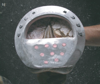
| Fig 8: Note the separations between the toe and heel quarters (red arrows). Separations removed using a rasp at a steep angle; trimming to a solid sole-wall junction. |
Farriery
Farriery will form the basis for treating WLD. The spectrum of WLD can range from a subtle hoof wall separation to an extensive separation which may cause substantial disruption of the stratum internum. Disruption of the laminar bond allows the distal phalanx to become unstable or to displace within the hoof capsule which would be consistent with mechanical laminitis. The treatment and farriery for chronic laminitis are beyond the scope of this review paper and will not be considered here.
Improving hoof conformation and correcting any hoof capsule distortion that may have contributed to the hoof wall separation is the initial step in treating WLD. In order to prevent small separations from progressing, the clinician should examine each foot carefully during routine trimming. Separations involving the inner hoof wall should be explored and debrided down to solid horn whenever possible and any cavity that is left after debridement should be removed. If there are solid hoof structures on either side of the separation, the separated area is removed by using the rasp at steep angle and trimming the outer wall down to a solid sole-wall junction (Fig 8). If the separation is considered too deep or extensive, it should be debrided as much as possible and then filled with a mixture of oakum, venice turpentine1 and copper sulphate or a medicated hoof putty2 before the solar surface of the foot is covered with a shoe. Filling the defect with copper sulphate may have some form of antimicrobial effect and filling the void under the shoe appears to allow the separation to grow down without further deterioration of the hoof.
Treatment of WLD must include protecting and unloading the damaged section of the foot with the appropriate farriery combined with resection of the detached hoof capsule overlying the affected area. A hoof wall resection will disrupt the continuity of the weight-bearing surface of the hoof capsule. Therefore, the authors feel that some type of shoe or device should be applied to restore the continuity of the weight bearing surface, to stabilise the hoof capsule and protect the resected area. Furthermore, the application of a shoe will prevent the horse from utilising the sole for weightbearing.
If the separated area of the hoof is determined to be extensive, it is important to initiate a farriery plan and then apply the appropriate farriery prior to resecting the outer hoof wall overlying the separation. Abnormal hoof conformation such as a long toe and or low heel, clubfoot and sheared heels is thought to contribute to the formation of WLD and should be addressed in the overall farriery plan. The farriery principles used to treat the hoof wall separations are to redistribute the load away from the defect, to unload the affected area and remove any leverage on the separated area.
 |
 |
 |
| Fig 9: Support shoes used to redistribute the weight away from the hoof wall. Wooden shoe (a), heel plate shoe (b) and heart bar shoe (c). |
The farriery always begins with the appropriate trim. As most separations occur in the dorsal hoof wall and extend to either the lateral or medial quarter, the weight-bearing is redistributed to the palmar/plantar foot. This can be accomplished by drawing a line across the widest part of the foot and trimming the hoof wall from this line palmarly/plantarly until the hoof wall and the frog are on the same horizontal plane. The trim increases the surface area in the palmar/plantar section of the foot recruiting it into ‘load sharing’ and generally creates two planes on the solar surface of the foot which will marginally unload the dorsal section to the foot (O’Grady and Poupard 2003). Excessive toe length, flares or leverage is reduced by trimming (rasping) the outer dorsal section of the hoof wall (Fig 5).
The type of shoe used and the method of attachment depend on the extent of the damaged hoof wall. If the defect is small, the hoof can be shod with an open shoe paying strict attention to any abnormal hoof conformation. If the defect is large and the overlying segment of detached hoof wall needs to be resected, some type of bar shoe is indicated to stabilise the hoof capsule. Often the shoe will need to be modified due to the loss of healthy hoof wall necessary for attachment. This can be accomplished by reshaping the shoe, punching additional nail holes in the shoe that approximate areas of solid horn and by adding clips to help secure the shoe. The shoe is fitted such that the branches of the shoe extend beyond the bearing border of the foot to provide additional weight-bearing surface in the palmar section of the foot. Breakover is placed palmar to the margin of the trimmed dorsal hoof wall to remove any leverage at the toe. This will also stop the ‘pinching’ effect that often occurs at the junction of normal hoof wall and detached horn at the proximal extent of the separation during breakover.
If the resection is to be extensive and/or if there is displacement of the distal phalanx, some type of supportive shoe or a wooden shoe should be used. Some form of supportive shoe allows some of the weight-bearing function to be transferred from the hoof wall to the frog (heart bar shoe) or to the frog, sole and bars (heel plate shoe) or the entire solar surface of the foot (wooden shoe) (Fig 9). Alternatively, the foot may be shod with an open shoe as described above and the solar surface of the foot between the branches of the shoe is filled with some type of silastic material (Fig 10).
 |
| Fig 10: Aluminum shoe with silastic material inserted between the branches to redistribute the weight-bearing on the solar surface of the foot. |
If the clinician is not comfortable using a traditional shoe, if there is limited hoof wall available in which to place nails or shoes cannot be nailed on safely, glue-on shoes may be another option (O’Grady and Watson 1999). Direct or indirect gluing can be utilised. Direct gluing attaches the shoe directly to the solar surface of the foot whereas indirect gluing attaches the shoe to the outer surface of the hoof wall using some type of cuff or plate. With either direct or indirect glue-on shoes, the resected area can be left exposed for treatment (Fig 11).
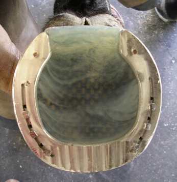
Recently, the first author has been using a wooden shoe on WLD cases with extensive hoof wall separations. The wooden shoe redistributes the load (weight of the horse) or reduces the forces on a section of the ground surface of the foot. This is accomplished by placing one flat solid surface against the solar surface of the foot. Breakover can be created in the shoe at any point around the perimeter to reduce leverage on the wall. The shoe is attached with small thin screws placed specifically in healthy wall, and the resected area can be left exposed for treatment (O’Grady and Steward 2009; O’Grady 2019) (Fig 12).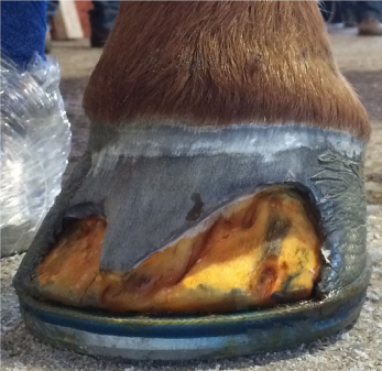 | 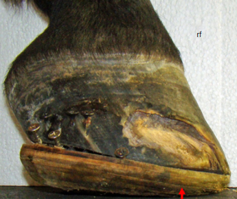 |
| Fig 11: Glue-on shoe using the indirect method of attachment. Using a cuff, the adhesive is only in contact with solid/healthy outer hoof wall, which eliminates the necessity for nails and there is no adhesive on the solar surface of the hoof. Note the exposure of the hoof wall resection for continued treatment and observation. Also, note the solid borders created around the periphery of the resection. | Fig 12: Wooden shoe attached with screws. Note the dorsal section of the foot unloaded and the breakover moved palmarly (red arrow). After applying 2-in casting tape, the resected area will still be exposed for treatment. |
If no rotation or displacement of the distal phalanx is present, a good ‘rule of thumb’ is that if greater than 1/3 of the overall surface area of the hoof wall is resected proximally, use palmar/plantar support. Foot casts and various types of boots have become popular in treating WLD especially after a resection has been performed; however, in the authors’ opinion, casts should be avoided as they tend to cover the affected section of the foot and boots create a continuous moist environment.
Resection
Complete hoof wall resection and debridement of all tracts and fissures in the affected area is necessary. This can be readily accomplished using a motorised Dremel3 tool with a tungsten carbide bit, a loop hoof knife and half-round hoof nippers. The debridement should be continued proximally and marginally until there is a solid attachment between the hoof wall and external lamellae (Fig 13). The clinician should not generally reach blood during debridement.
 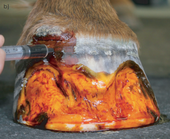 |
| Fig 13: Hoof wall resections. It is important to create and maintain around the perimeter of the resections solid margins noted on both feet in this illustration. Maintaining a solid junction prevents the separation from continuing proximally. Dye marker (2% tincture of iodine) being applied to the resection to show up the tracts in stratum internum. These tracts will generally require further debridement at the next recheck. |
Topical disinfectants and medications in any form are of no value without resection of the affected hoof wall. A plethora of topical medications have been described for treatment following hoof wall resection but there have been no controlled studies on any product, and none have any proof of efficacy. Antiseptics/astringents such as Merthiolate or 2% iodine are commonly used; however, their most beneficial effect may be as a dye marker to outline any remaining tracts (Fig 13). The dye marker will serve as an aid in making the remaining tracts more visible at subsequent examinations and as a guideline during subsequent debridement. After thorough hoof wall resection, the affected area can be left open to grow out with debridement at frequent intervals. The authors use a wire brush daily to keep the resected area clean (O’Grady 2002; O’Grady 2006; Pleasant and O’Grady 2008; O’Grady 2011). Following the initial debridement, thorough exploration and debridement of any remaining tracts should take place at 2- week intervals by the veterinarian or farrier. When the resection has grown out, a thorough examination of the sole/ wall junction is imperative at re-shoeing intervals of 4– 5 weeks.
Complications
The authors feel that if an extensive hoof wall resection is performed in one or multiple feet, the horse should be taken out of work. A composite (acrylic or urethane) repair of the resected area is often requested by the client to keep the horse in work but this practice should be discouraged. An acrylic (poly methyl-methacrylate) material4 with or without an antibiotic is still commonly used to repair a hoof wall resection associated with WLD (Turner and Anderson 1996). When an acrylic hoof repair material impregnated with a powdered antibiotic is used, it requires moisture against the composite for the antibiotic to exude from the acrylic. Furthermore, the acrylic composite cures with marked hyperthermia which can create moisture under the repair.
Finally, any organisms on the surface of the stratum internum will be sealed under the repair and the heat generated when the acrylic cures make the underlying tissue softer, more permeable and may allow organisms entry into the dermis. Therefore, using an acrylic repair is counterintuitive to the accepted practice of creating a dry cornified surface of the stratum internum and allowing it to grow out. A repair should only be considered in selected cases where the client is unable to treat the resected area and where cosmetics are a necessity such as when a horse is competing in a nonperformance class. In this case, if a repair is applied, there should be a significant interface (silastic material or clay) placed between the composite and the stratum internum. In all cases, composites may hide and/or foster infection and tend to weaken the surrounding solid hoof wall, all of which can encourage continued propagation of a hoof wall separation or re-infection (O’Grady 2002; O’Grady 2006; Pleasant and O’Grady 2008; O’Grady 2011; Redding and O’Grady 2012).
Burr holes
The use of creating a burr hole through the hoof wall at the proximal extent of the separation has been described in farrier and lay publications. The rational behind this procedure is to attempt to treat the separated area by flushing with some type of medication without removing the overlying hoof wall. It is further thought that by treating WLD in this manner and not resecting the detached hoof wall, the horse can stay in work. However, the authors feel that if the separation is that extensive to warrant this type of treatment, the foot is compromised, the distal phalanx is unstable and has the potential for further damage. Furthermore, flushing does not remove the degenerating horn present within the separation and maintains a moist environment under the hoof wall.
Aftercare
A change in environment is important. The feet should be kept as dry as possible throughout the recovery period. Sawdust or wood shavings appear to dehydrate the feet making them the bedding of choice; the bedding should always be kept clean and dry. Limited turnout in rain or wet weather is helpful. Turnout can be delayed in the morning until the sun has dried the dew from the pasture.
A commitment from the owner with regards to a continuous treatment schedule is essential until all signs of disease have been eliminated, and the hoof wall is replaced with new growth. Equally important is maintaining a consistent schedule with the farrier who will continue the appropriate farriery and monitor the resection as it grows out. The extent of the damage will determine the approximate amount of time required to complete the treatment process.
Results
There are many contributing factors to consider when assessing the results of treating WLD. These factors would include an early and accurate diagnosis, the extent of the hoof wall defect, understanding the abnormal forces placed on the foot, the application of the appropriate farriery and the importance of owner compliance. Early recognition and treatment of WLD along with the use of frog and sole support materials is essential in order to prevent displacement of the distal phalanx. Obviously, the results would be further complicated if a case of WLD is associated with a marked displacement of the distal phalanx within the hoof capsule. The success of the authors’ treating of WLD may be somewhat biased as both their practices are referral practices limited to the equine foot. The records from the authors’ combined practices on greater than 100 cases presented with extensive WLD were reviewed. The treatment protocol was similar in both authors’ practices: consisted of application of the appropriate farriery, resection of the devitalised hoof wall and owner using a wire brush on the resected area daily. In some cases, a dye marker was used to aid in recognising tracts in the epidermal lamellae on subsequent debridements. Follow-up on 70% of the cases showed complete resolution of the disease process. The other 30% of the cases were lost to follow-up or lack of compliance from the caretaker with regards to continuing treatment/ farriery.
Prevention
Prevention of WLD is difficult because the exact aetiology is unknown. Discussing the potential problem with the farrier and having them examine each foot when the horse is shod is extremely important and leads to early recognition. Any small abnormal separation or defect involving the inner hoof wall adjacent to the sole-wall junction should be noted, explored and debrided down to solid horn. Good basic farriery is essential for creating and maintaining a strong sole/ wall junction that may prevent separations and offer protection (O’Grady and Poupard 2003). Equally important is the necessity to carefully monitor horses that have previously had WLD as they tend to reoccur. A year or two after WLD has been successively treated and resolved, it can suddenly reappear in some horses with strong hoof walls that have shown no previous signs of a hoof wall separation.
Discussion
White line disease involves the inner, nonpigmented section of the stratum medium of the hoof wall, not the sole-wall junction (zona alba, or ‘white line’). Thus, ‘white line disease’ is somewhat of a misnomer; however, it has become the accepted term used by most farriers and veterinarians. Certainly, it is a more useful term than onychomycosis, which implies a human disease and limits the primary aetiological organism to a fungal agent.
Treating WLD is often a dilemma for owners, veterinarians and farriers. Owners have been deluged with many proposed causes and a variety of treatment protocols. Numerous commercially available preparations have been marketed for treating WLD, all claiming success. The internet and social media describe a multitude of products and methods guaranteed to provide miraculous improvement. However, presently, there is no convincing scientific evidence as to the efficacy of any given product on the market. Veterinarians are often unaware of the magnitude of this problem or potential consequences of WLD as they only see the severe cases that present with a marked hoof capsule distortion, for lameness evaluation or when radiographic changes become apparent. However, WLD may be a subtle contributor to hoof capsule disorders such as a change in shape, full-thickness hoof wall cracks or abscessation; additionally, it may lead to other causes of lameness within the foot such as laminitis. Farriers generally see horses on a routine basis and need to be in the forefront regarding treatment and especially prevention of WLD. They need to be aware of hoof wall separations encountered during routine farriery (trimming and shoeing) and realise these separations have the potential to advance and extend proximally and marginally in the hoof wall. Abnormal hoof capsule conformation associated with hoof wall separations such as a long toe, clubfoot or sheared heels should be recognised and addressed. Farriers are aware of this disease as they are often confronted with nailing a shoe on to a limited or compromised hoof wall and keeping the shoe on between resets. Farriers often resort to using topical treatments since owners are reluctant to have resections performed; many farriers are reluctant to recommend resections – a procedure that can be daunting and that takes the horse out of work. Research, continued veterinarian/farrier awareness of WLD along with owner education appear to be the most promising direction for the future. There is ample evidence in this paper and other work that traditional farriery is effective in treating and resolving WLD. Research should look at the pathophysiological mechanism where a minor hoof wall separation advances to a marked pathological disease process. Understanding this pathway would provide additional information as to how WLD could be prevented. Quality continuing education (CE) would alert both veterinarians and farriers to the significance of hoof wall separations and their potential to progress to WLD. Furthermore, it appears that abnormal hoof capsule conformation or distortions are risk factors for WLD; therefore, educating both veterinarians and farriers to recognise and address these distortions with appropriate farriery would be useful. The successful management of WLD demonstrates the importance of maintaining and promoting a functional veterinarian–farrier relationship.
ACKNOWLEDGMENTSAuthors’ declaration of interests
No conflicts of interest have been declared.
Ethical animal research
Not applicable to this review article.
Authorship
S. E. O’Grady did the literature search. S. E. O’Grady and T. D. Burns used their combined clinical cases for reviewing the consistent, successful treatment of a large number of white line disease cases. S. E. O’Grady and T. D. Burns revised the manuscript.