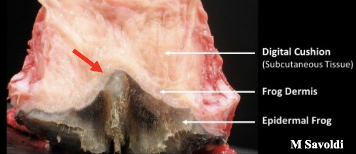
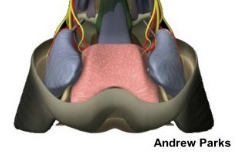
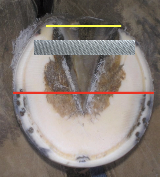
| Farriery for the Equine Frog / Digital Cushion Complex Stephen E. O’Grady, DVM The importance of the equine frog regarding the health and function of the foot is often not recognized. Furthermore, it is hard to divorce or separate the frog from its association with the digital cushion (Figure 1). The conformation of the palmar / plantar section (and the overall conformation) of the foot is determined by the size and mass of these combined structures. Furthermore, there appears to be a symbiotic relationship between these two structures such that when the size of digital cushion decreases, the mass of the frog will increase due to additional forces. Farriery plays a major role in maintaining the health and destiny of these two structures. The different components of the palmar foot, including the laminar junction, hoof wall, bars, ungual cartilages, frog, and digital cushion; each play a significant role in dissipation and distribution of force (Figure 2). Any dysfunction in one or all these components of the palmar foot will lead to forces being transferred to more sensitive structures, either within the hoof itself or to more proximal structures, causing subsequent injury. Two theories have been proposed to explain the mechanism of force distribution using the soft tissue structures in the palmar foot. The pressure theory suggests that the frog pushes upward into the digital cushion at ground contact to force the lateral cartilages outward. On the other hand, the depression theory emphasizes the downward movement of the pastern into the digital cushion, which in turn forces the lateral cartilages outwardly. However, neither theory can explain the large negative pressure present within the deep areas of the digital cushion during both stance and locomotion (Dyhre-Poulsen P, et al. EVJ 1994). Considering the structural organization of the lateral cartilages, digital cushion, and enclosed vasculature, this suggests the likelihood that a hemodynamic mechanism plays a significant role in the dissipation and distribution of force during the landing and stance phase of the stride. However, the exact mechanism of force distribution and anti-concussive properties of the palmar foot is not fully understood and will not be debated here.
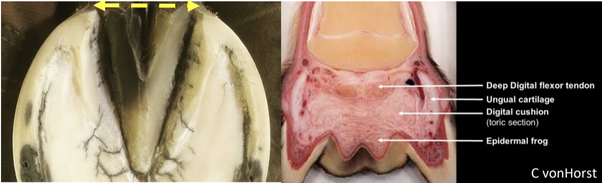
Anatomy The frog (cuneus ungulae) is a wedge - shaped mass of keratinized stratified squamous epithelium with a softer structural texture because of an increased moisture content. Spherical masses of apocrine glands present in the corium of the frog have ducts that deliver secretions to the surface of the frog. This further contributes to the rubbery nature of the frog. The ground surface of the frog presents a pointed apex and central sulcus enclosed by two crura. Paracuneal (collateral) sulci separate the crura of the frog from the bars and the sole. The palmar aspect of the frog blends into the bulbs of the heels (Figure 3). The digital cushion (pulvinus digitalis) is a highly modified subcutis consisting of a meshwork of collagenous and elastic fibers, adipose tissue, and small bundles of fibrocartilage. The digital cushion lies just proximal to the frog and is fixed to the adjacent structures by ligaments and thick fiber bundles. Only a few blood vessels ramify in the digital cushion. It is divided into two parts; the large bilobed toric part of the digital cushion fills the bulk of the space between the heels and bulges palmarly to fill out the bulbs of the heels. The cunean part is a smaller v-shaped collagenous extension of the toric part underlying the apex of the frog (Figure 4). The cells that comprise the digital cushion are slow dividing and as only a few blood vessels ramify in the structure, therefore; it is extremely unlikely that it can restore or increase in size.
Function Function is based on the frog and digital cushion working in unison. As part of the epidermal hoof capsule, the frog would surely play a role in protection for the internal structures located above. Traction would be another function where the base of the frog and the descent of the apex would act as a brake on soft footing or on a slippery surface. Obviously, some of this traction would be assumed by the sharp edge of the shoe at the toe when shod. Together with the digital cushion, the complex plays a major part in the dissipation of the energy of impact and anti-concussion during the stance phase of the stride. Finally, an overlooked function of the frog is to act as an expansion joint to maintain the width of the heels. If the frog becomes recessed and loses mass or C vonHorst if the heels migrate dorsally, the heels of the hoof capsule will contract (Figure 5). The frog and digital cushion provide a deformable interface between the rigid structures of the palmar hoof capsule that permits smooth expansion and recoil during weight bearing. Although the digital cushion is a predominant factor in absorbing the energy of impact with the ground, it must be combined with other soft tissue structures such as the coronary cushion, the laminar junction, the lateral cartilages and the extensive network of venous plexuses throughout the foot which are involved in this complicated function. From a straightforward mechanical perspective, a large frog and a well-formed palmar foot will play a significant role in cushioning hoof impact which argues for the necessity of maintaining a large strong healthy frog /digital cushion complex.
Farriery The appropriate farriery is essential to maintain or promote a healthy frog. Furthermore, it is extremely rare to see thrush when the frog has adequate size and substance. At the onset of the trim, it is easy to assess the dimensions of the frog either by measurements or just visualization. The width of a healthy frog should be 65-70% of its length (Turner 1988). The frog should not be excessively recessed between the heels of the hoof capsule or protrude below the ground surface of the heels. The frog will generally be slightly recessed at the end of a 4-6-week shoeing cycle as the heels grow forward during this time. The author’s basic approach to trimming the palmar foot will be outlined here. A line is drawn or visualized across the widest part of the foot, the frog and sulci are cleaned with a wire brush and any loose exfoliating horn is removed from the frog. The surface at the base of the frog is reduced if it is slightly protruding below the ground surface of the foot. The heels of the hoof capsule are trimmed palmarly / plantarly to where the hoof wall and the frog are on the same plane. This will usually extent the ground surface of the heels to the base of the frog and places the heels and frog in a ‘load sharing’ position. Frog pressure has and will be debated endlessly, however, it’s the author’s contention that when a flat shoe is placed on the horse’s foot, the descent of the frog from the weight of the horse counteracted by the deformable surface which the horse usually encounters, adequate but not excessive pressure is placed on the frog.
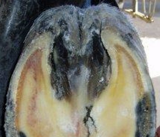
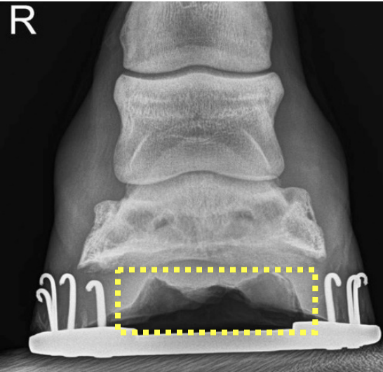
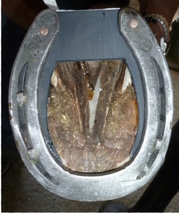
Abnormal Hoof Conformation and the Frog It is a physiological fact that the palmar foot should be loaded but no individual component should be overloaded. Hoof capsule distortions are among the varied causes that change the size, shape, and position of the frog. The change in position, either recessed or protruding below the ground surface will always be accompanied by a change in size and shape. When the frog is recessed between the heels of the hoof capsule, the expansion joint is lost, the heels will narrow and contract, and the load bearing will be directly shifted to the hoof wall. Furthermore, the void left from the decreased size of the frog will fill with dirt and debris, now; instead of distributing the load it will put excessive pressure on the compromised frog and damage it further (Figure 7). It also creates the ideal environment for thrush. This void left by the diminished size of the frog can be easily noted when veterinarians and farriers are evaluating radiographs (Figure 8). A recessed frog often occurs in upright feet or when a flexural deformity is present.
Generally, the heels of the hoof capsule and the frog can be aligned during routine trimming as described above and must be combined with the appropriate size shoe. When a flexural deformity (club foot) is present, the frog will be markedly recessed between the hoof wall which makes the task of putting the frog and hoof wall on the same plane harder as there is a shortening of the deep digital flexor muscle tendon unit. However, the palmar foot still needs to be loaded. In this case, the heels are trimmed to the plane of the frog to load the foot, toe length is reduced, and mild heel elevation is applied to compensate for the decreased length or shortening of the DDF muscle tendon unit (O’Grady 2012) (Figure 9A & 9B).
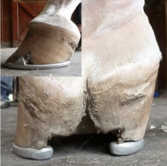
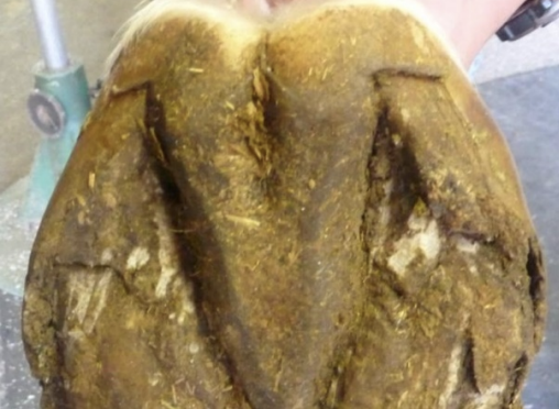
The other scenario is the frog protruding or prolapsing below the ground surface of the foot and is generally seen with the long toe and or low heel hoof capsule distortion. It can be caused by damage to the digital cushion or the heels of the hoof capsule migrating dorsally such that the frog will displace palmarly and downward. Over time, the frog will become large and bulbous due to stimulation from the ground and to counteract excessive weight bearing. The heels will now have to be trimmed using the rasp alongside of the frog instead of using it across the palmar section of the foot. In the reverse situation from the recessed frog, more weight bearing is transferred to the protruding frog and decreased on the hoof wall. There also appears to be a correlation in horses with low heels, that when the mass of the digital cushion decreases from overload, the mass of the frog increases to compensate (Figure 10A & 10 B).
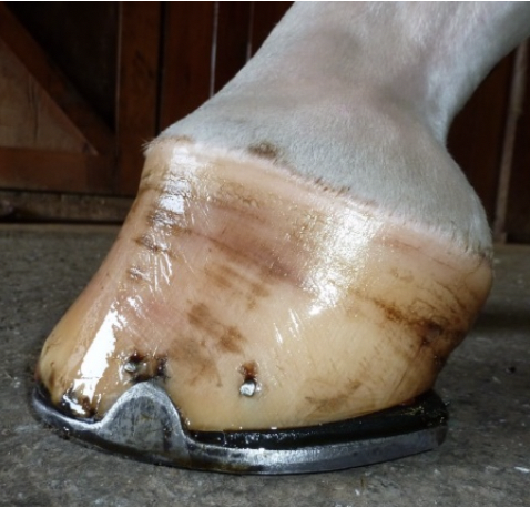
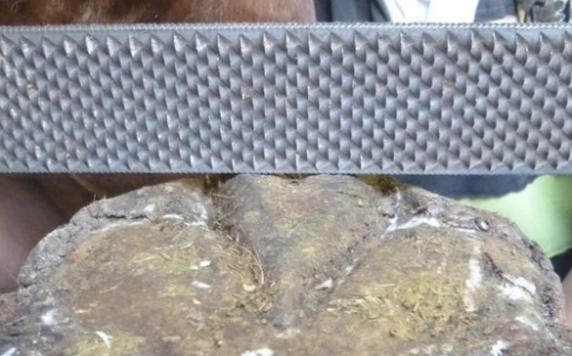
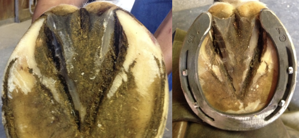
The object of the farriery here is to reduce the amount of frog below the ground surface of the foot and return the palmar foot to a’ load sharing’ function. Obviously, the method used is dependent on the size of the frog and duration. Changing the position of the frog can be accomplished in many ways. A wooden shoe can be applied briefly which will quickly put the heels and frog on the same plane (also improve a thin sole). Using a shoe with a frog or heel plate, which puts direct pressure on the frog, will decrease its size and allow the heels to assume a better position. If time permits, one of the author’s favorite methods is to remove the shoes and briefly leave the horse barefoot. The horse is placed on a walking program until the frog is reduced to the same plane as the hoof wall. The heels are now trimmed at 10-day intervals until the palmar foot is load sharing which usually takes 6-8 weeks. The shoes are then replaced (Figure 11A &11B).
The exact mechanism by which the horse loads its foot is unclear and continually being studied. However, when the foot strikes the ground, the distal phalanx descends distally and palmarly/plantarly causing the quarters to flare and move outwardly. It appears that the main contributor to heel motion is a continuation of this flaring of the quarters during weight bearing (Thomason 2007). In the palmar foot, all of the soft tissue structures are important in absorbing energy when the foot impacts the ground and as the frog /digital cushion complex form the bulk of the soft tissue; maintaining the health of this component of the palmar foot is essential. | ||||||||||||||||||||||||||||