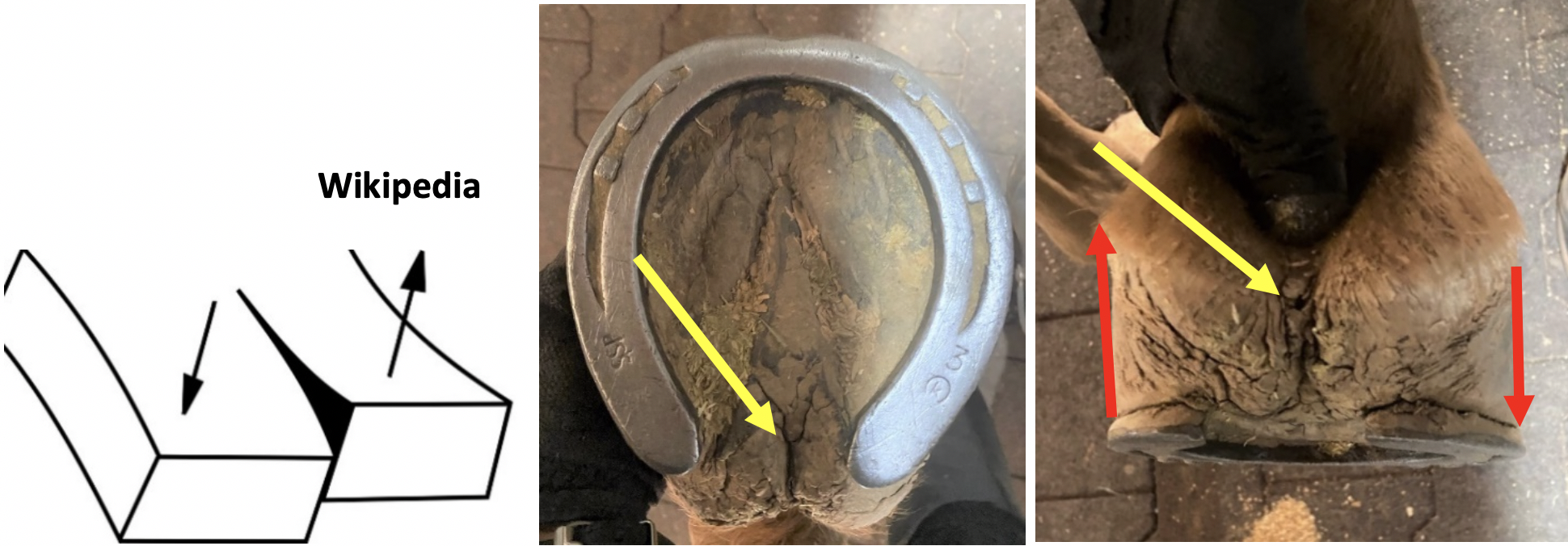
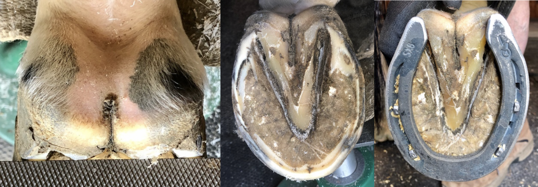
| Fissure Formation at the Base of the Frog Stephen E. O’Grady, DVM A fissure or split occurring through the central sulcus of the frog has always been characterized as thrush in both the veterinary and farrier literature along with a myriad of proposed treatments. There is little doubt that thrush does not occur secondarily to the formation of a fissure; however, in the author’s opinion, the primary cause of the fissure or separation in the frog tissue is due to parallel shearing forces exerted on the hoof wall in opposite directions. The fissure will occur between the two forces in the weakest part of the soft tissue (Figure 1). Like thrush found in the body and sulci of the frog, a fissure at the base of the frog is rarely seen in a healthy frog. This condition can be seen in horses with any form of hoof capsule distortion, a compromised frog, and combined with an asymmetrical strike pattern when the foot lands on the ground (Figure 2). The fissure will often extend into the skin above the bulbs of the heels causing bleeding and a source of lameness.
The formation of a fissure in the central sulcus of the frog is straightforward. It begins when the frog loses functionality which also sets up the potential to acquire thrush. The fundamental problem with palmar/plantar conformation usually involves the frog not being near or on the same plane as the heels, thus not having contact with the ground causing the frog to atrophy. This can occur for a variety of reasons, including many of the recognized hoof capsule distortions such as the low heel, clubfoot, or sheared heels along with inappropriate farriery. An unhealthy frog is markedly smaller in size and recessed below the level of the ground surface of the hoof, thus creating a void in which debris can accumulate. It appears that as the frog loses size and mass, the structural mass of the digital cushion also decreases. As the frog recedes within the hoof capsule, the frog loses its “self-cleaning mechanism,” which allows material (dirt, manure, etc.) to accumulate over the frog, creating excessive pressure. Over time, this constant pressure on a compromised frog leads to increased deterioration and further atrophy. Weakened by the reduced protective horn of the epidermis, the frog tissue Wikipedia becomes susceptible to penetration by bacteria and other organisms leading to the development of the thrush. The frog / digital cushion complex forms the bulk of the palmar foot and forms the connection between the heels of the hoof capsule. An asymmetrical landing pattern as noted in sheared heels results in an unequal force placed on each heel, causing the heels to move in opposite directions. This movement in opposite directions causes shear force localized to the weakest part of the soft tissue, which is the central sulcus of the frog to separate or tear forming a fissure. Significant movement in the fissure can often be demonstrated by manually moving the heel bulbs in opposite directions (Figures 3A, B & C).
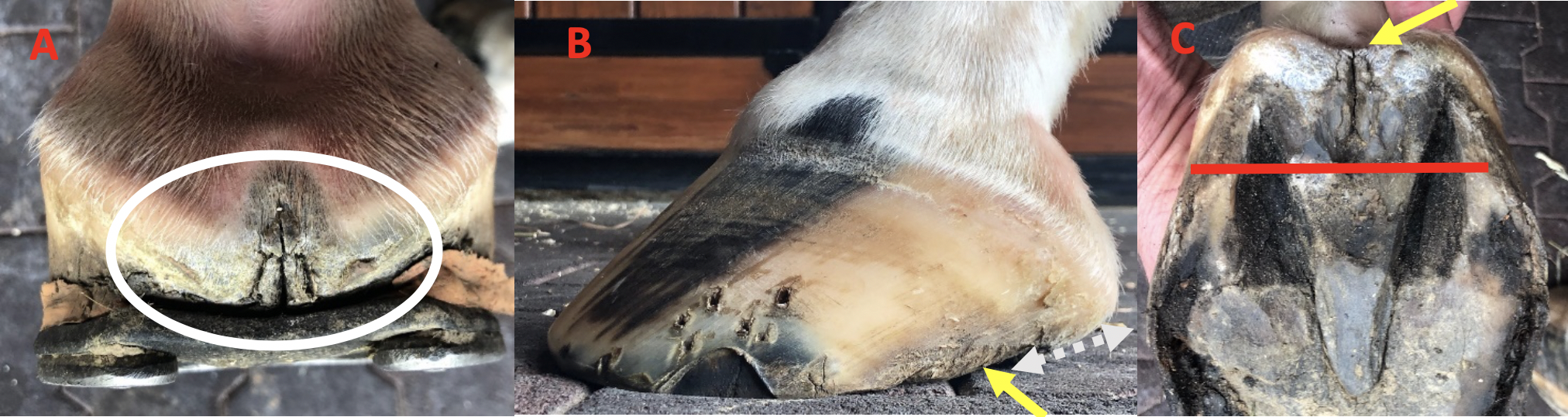
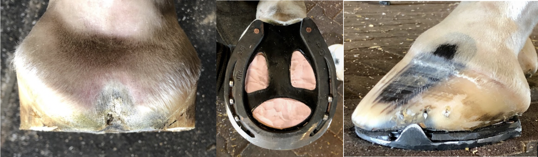
A frog fissure also be seen with low heel conformation. In this case, the heels migrate dorsally while the soft tissue structures move or prolapse palmarly / plantarly and are no longer housed within the hoof capsule. With this conformation, the frog and digital cushion generally show a decreased mass, are no longer supported, placed under stress, and prolapse toward the ground. Now, with the heels migrated dorsally and the soft tissue palmarly…the soft tissue is unstable and subject to a shear force (Figure 4A, B & C).
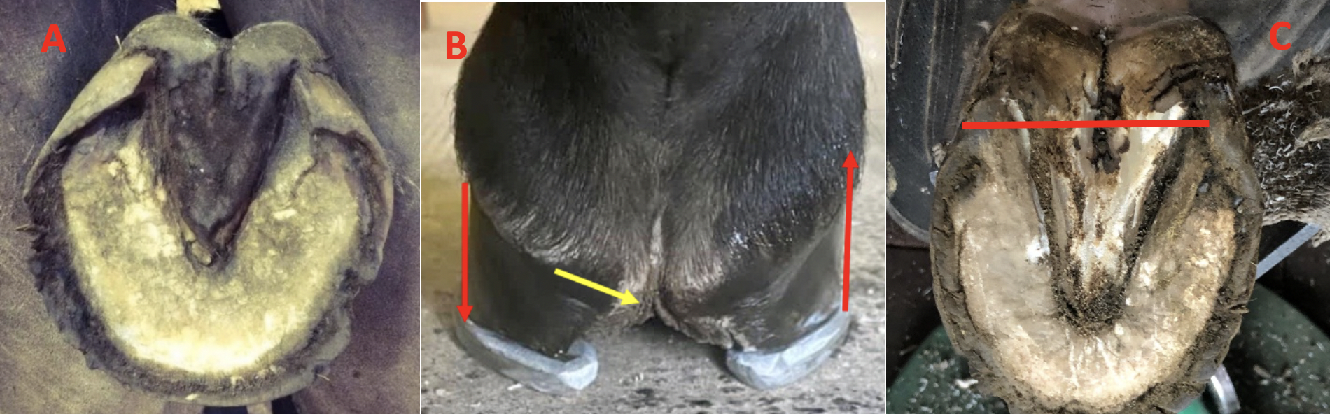
The fissure will also readily occur with a low/under run heel foot conformation (Figure 5A). Horses with a clubfoot (flexural deformity) or an upright foot will produce excessive heel to compensate for the shortened length of the DDF muscle tendon unit. With the increased heel height, the frog will often be recessed between the hoof wall, undergo atrophy, and loses structural mass. This leaves an unstable soft tissue link between the heels and the independent movement of the heels sets up a shear force at the weakest point of the frog which is the central sulcus (Figure 5B). A fissure will also readily occur in the hindfeet as well when a low heel ‘bullnose’ conformation is present. Here again the heels of the hoof capsule can migrate forward, and the frog/digital cushion prolapse plantarly which can cause a shear force in the soft tissue (Figure 5C).
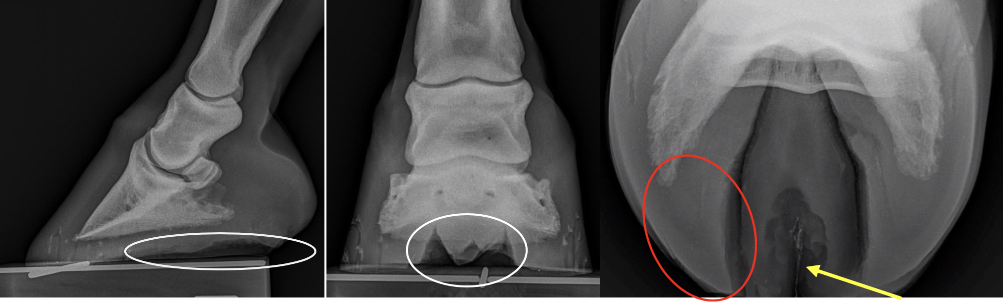
The structure of the frog and the frog fissure can readily be observed when evaluating foot radiographs. On the lateral and DP radiograph, a lucency can be seen which indicates the frog has recessed between the heels of the hoof capsule. On the tangential or skyline view, the outline of the frog, the frog fissure and the structures of the heels can be examined (Figure 6). Bearing in mind, that in a healthy frog, the width of the frog should be 70% of its length (Turner 1988). Furthermore, the heels act as an expansion joint between the heels; therefore, when the frog narrows, the heels move axially and contract. The heels of the hoof capsule take on an appearance that resembles the ‘head of a bird’ which can be viewed on the radiograph.
Farriery Farriery will be directed at improving the structure of the frog, improving hoof capsule conformation that led to the unhealthy frog and stabilizing the heels of the hoof capsule. Please see here for an overview of the equine frog and its farriery. The first step is to make the heels ‘load sharing’ and this is accomplished by trimming the heels of the hoof capsule to the same plane as the frog. This can be accomplished in most cases and it immediately decreases the shear in the soft tissue and adds stability. After trimming, when possible, the horse is left barefoot for as little as 7-10 days in a stall with minimal bedding and hand walked. The longer the horse remains barefoot, the more improvement will be seen in the frog / palmar section of the foot. Please see here. The heels will relax, the frog will change shape, the fissure will decrease in length and frog will start to produce horn. Leaving the horse barefoot is extremely helpful when the frog is prolapsed below the ground surface of the foot as noted in Figure 4A as the weight of the horse will reposition the structures in the palmar foot. When ready to be shod, the foot is trimmed to address any distortions present and a shoe such as a bar shoe, a shoe with a heel plate or a shoe with a spider plate can be used to stabilize the heels and thus prevent or decrease the vertical movement. If the horse can have sufficient time barefoot and the fissure has improved in length and depth, an open heeled shoe may be appropriate (Figure 7A B & C).
Treating the fissure I have always questioned whether thrush is actually a localized infection as one would see in a wound. Perhaps we could consider thrush to be devitalized horn that gets contaminated with organisms found in the ground / envirnoment. Interestingly, when diseased frog is cultured, the same two opportunistic bacteria are always isolated. However, if we think about the frog fissure as a wound, the two factorss that will prevent or delay healing is infection and movement. Once the appropriate farriery has been applied and the heels are stabilized, I find very little topical treatment is necessary. After the horse is shod, I will generally soak the foot one time with a saturated solution of epson salts (MgSO4) to clean the fissure. If the fissue is deep, I will use betadine solution or sugardine (betadine soln. and sugar) in the wound and then pack the fissure with a strand of gauze. I have always had a rule that I never use any topical medication on a wound in the foot that I wouldn’t use on my own skin. If the fissure is not deep or as healing progresses, I will just pack the defect with dry gauze without medication. If the fissure is kept clean and dry, it will heal. If the fissure is deep and has been chronic, the horn will often heal in a involuted manner which will leave a shallow dry trough at the cental sulcus of the frog through the bulbs. I would like to recognize farriers Tyson Clark, CJF and Jeff Ridley, CJF, TE who participated in these cases, for their cooperation, insight and skilled farriery | ||||||||||||||||||||