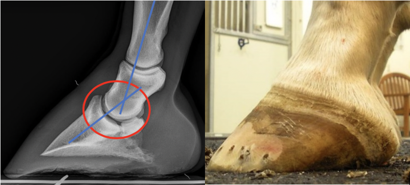
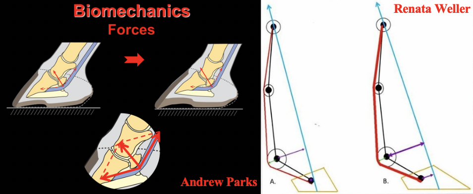
| The Negative Palmar Angle Stephen E. O’Grady, DVM The negative palmar angle (NPA) or more correctly, ‘the angle of the solar border of the distal phalanx’, has become a diagnosis, a disease, and a focus for a myriad of veterinary / farriery treatments. Simply stated, the negative palmar angle results from a loss of the soft tissue structures (especially the digital cushion) in the palmar section of the foot. The loss of mass allows the distal phalanx to descend distally in the palmar section of the hoof capsule. The result is a change in the position of the distal interphalangeal (DIP) joint which places the joint in dorsiflexion and a distortion of the hoof conformation. This is not to say that the NPA does not or cannot have negative consequences on soundness. The change of position of the bone creates a broken back hoof-pastern axis, changes the position of the (DIP) joint which changes the distribution of weight bearing on the solar surface of the foot and increases the tension in the deep digital flexor tendon (DDFT) (Figure 1). The decreased mass/density of the soft tissue structures in the palmar foot hinder the foot from dissipating the energy of impact and providing adequate shock absorption. The sling formed by the DDFT under the navicular bone descends into the soft tissue structures as the joint moves in a distopalmar direction when the heel strikes the ground, thus forming one of the initial shock absorbing mechanisms of the foot. This shock absorbing property is lost with a significant decrease in soft tissue mass (Figure 2). All these changes can have deleterious effects on the equine foot and soundness.
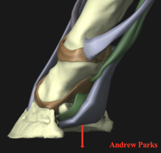
The biomechanical aspects and the correlation to lameness related to the effect of the negative angle of the distal phalanx on the podotrochlear apparatus (navicular area) is huge. Eliashar (Eliashar et al EVJ 2004) showed that a 1° decrease in palmar angle of the distal phalanx results in a 4% increase in peak force on the navicular bone. This places excessive load on the navicular bone, the navicular bursa, and the distal extent of the DDFT (Figure 3). Furthermore, the horse will now spend a substantial amount of time with the DIP joint in dorsiflexion which is not only an abnormal position of the joint but also changes the pattern of weight bearing on the solar surface of the foot.
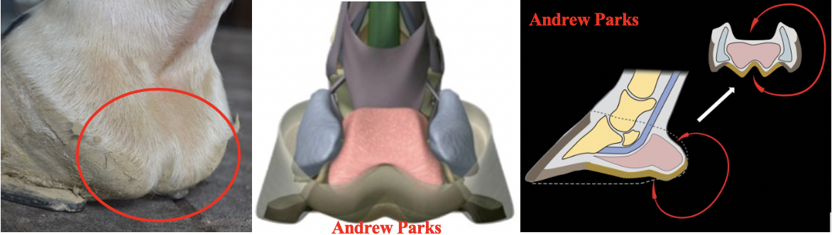
Improving hoof conformation to address the negative palmar angle of the distal phalanx presents clinicians with a farriery challenge. Often improvement cannot be achieved and therefore, the focus is to maintain the compromised structures. Traditional farriery methods have been to provide heel elevation to improve the digital alignment and reposition the DIP joint. However, this practice can be detrimental if the soft tissue structures in the palmar foot are compromised or lack the appropriate mass. When heel elevation is added, it temporarily improves the appearance of the hoof capsule, but the pressure placed on the soft tissue structures overtime will damage them further. The limiting factor when attempting to improve the structures in the palmar foot is the inability to grow hoof wall in the heel area necessary to improve the hoof capsule. The primary soft tissue structure involved in the compromised palmar foot is the digital cushion. Genetics are incriminated for the decreased size of the digital cushion as many animals are born (TBs for example) with inadequate tissue while other factors, in the author’s opinion are lack of development as a juvenile, training regimens, overuse, surfaces and farriery practices. Considering the cells that comprise the digital cushion, there is little evidence to suggest that it can restore or increase in mass. See here. When the mass of the digital cushion is decreased, more weight is placed on the frog which causes it to enlarge and often protrude or prolapse below the solar surface of the foot. It is the author’s opinion, based on many years of farriery, that hoof wall growth requires a structural framework between the heels of the hoof capsule to produce horn. Loss of that framework occurs when the mass in the palmar foot markedly is decreased (Figure 4).
Farriery for the negative palmar angle must be based on farriery principles such as protection of the compromised structures and redistribution of the forces to other areas of the foot. This condition requires careful evaluation of the foot conformation, amount of damage to the structures of the palmar foot, landing pattern of the horse, the surface on which the horse works and the appropriate farriery necessary to address the above principles. For further information on hoof capsule distortions see here.
|