How to Manage the Club Foot-Birth to MaturityReprinted with permission from the American Association of Equine Practitioners. |
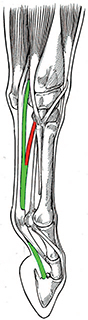 Fig. 1. |
| Fig. 1. Illustration shows the structures involved in a flexural deformity of the DIPJ. Note the close association between the AL-DDFT (red line) and the DDFT (green line). |
3. Classification of Club Feet
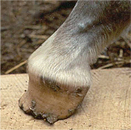 Fig. 2. |
| Fig. 2. Grade 4 club foot. Note the broken forward hoof-pastern axis, fullness of the coronet, the disparity between hoof wall growth at the toe and the heel, the concavity in the dorsal hoof wall, and the poor hoof wall consistency at the ground surface of the capsule. |
4. Club Feet in the Young Horse
Club feet or flexural deformities in foals can be divided into congenital or acquired deformities. As such, congenital deformities are noted at birth, and acquired deformities generally occur from 2 to 8 months of age as the foal grows and develops.2-4,10
Congenital Flexure Deformities
Congenital flexural deformities are present at birth, may involve a combination of joints (e.g., carpus, metacarpophalangeal, and distal interphalangeal joints), and are characterized by abnormal flexion of these joints and the inability of these joints to extend. Proposed etiologies of congenital flexural deformities include malpositioning of the fetus in utero, nutritional mismanagement of the mare during gestation, teratogens in various forages ingested by the mare, maternal exposure to influenza virus, or the deformities could be genetic in origin.2,6,8,10 The affected foal tends to walk on the toe of the hoof capsule, is unable to place the heel on the ground, and assumes a so-called "ballerina" stance. Treatment of foals with a congenital flexural deformity varies with the severity of the deformity. A mild to moderate flexural deformity in which the foal can readily stand, nurse, and ambulate is generally self limiting and resolves without treatment. Brief intervals of exercise once or twice daily in a small paddock or on firm footing for the first few days of life may be all that is necessary for the deformity to resolve. If the condition is severe or has not improved by the third day post-foaling, every-other day intravenous administration of oxytetracycline (2-3 g q 24 hrs) is frequently beneficial.2,10,11 A variety of bandaging techniques, often combined with splints, can be used to fatigue the muscle portion of the musculotendinous unit. Physical therapy to "stretch" the involved area may hasten recovery. Foals with bilateral congenital flexural deformities usually don't have just 1 isolated structure or joint that is responsible for the deformity, therefore, in the author's opinion, the use of a toe extension is not indicated. In the author's experience, a toe extension will often impede movement and cause the foal to stumble when it attempts to ambulate.
Acquired Flexural Deformities
Acquired flexural deformities generally develop when the foal is between 2 and 8 months old and generally involves the DIPJ initially. It is commonly a unilateral condition but occasionally affects both limbs. The etiology of this deformity is unknown, but speculated causes include genetic predisposition, improper nutrition (i.e., overfeeding, excessive carbohydrate [energy] intake, unbalanced minerals in the diet), and excessive exercise.2,10 A recent study looking at grazing patterns in a small number of foals showed that foals with long legs and short necks had a tendency to graze with the same limb protracted.12 Fifty percent of the foals in this study developed uneven feet with a higher heel on the protracted limb leading researchers to feel there may be a possible correlation between conformational traits and an acquired flexural deformity. It is the author's opinion that a large contributing factor to this syndrome is contraction of the muscular portion of the musculotendinous unit caused by a pain response, the source of which could be discomfort anywhere along the limb, physeal dysplasia, or trauma from foals exercising on hard ground. Discomfort may follow aggressive hoof trimming where excessive sole is removed, thus rendering the immature structures within the hoof capsule void of protection. The foal then becomes unwilling to bear full weight and is susceptible to trauma and bruising. Any discomfort or pain in the foot or lower portion of the limb coupled with reduced weight bearing on the affected limb appears to initiate a flexor withdrawal reflex; this causes the flexor muscles proximal to the tendon to contract, leading to a shortened musculotendinous unit and an altered position of the DIPJ. This shortening of the musculotendinous unit shifts weight-bearing to the dorsal section of the foot causing a decrease in sole depth, bruising of the sole, reduced growth of the dorsal aspect of the hoof wall, and excessive hoof wall growth at the heel. As the flexural deformity may be secondary to pain in these cases, it is essential that a possible source of pain should be carefully evaluated and located by physical examination and, if necessary, by regional analgesia and diagnostic imaging. A genetic component must also be considered for acquired flexure deformities, as some mares consistently produce foals that develop a flexural deformity in the same limb of the dam or grand dam in which a similar deformity is present.10 However, at present, there is no conclusive scientific evidence to substantiate a genetic basis.
5. Mild Acquired Flexural Deformities (Initial Stage of a Club Foot)
Clinical Signs
The initial clinical sign of a club foot may only be abnormal wear of the hoof at the toe, which is often discovered by the farrier during routine hoof care. Closer or subsequent investigation may reveal that the dorsal hoof wall angle is increased and that after the heels of the hoof capsule have been trimmed to a normal length, the heels may no longer contact the ground. A prominent coronary band may be present at this stage. Most foals affected to this degree may already have a mildly broken forward hoof pastern axis. Increased palpable digital pulse, heat in the affected foot, and signs of pain when a small hoof tester is applied to the solar aspect of the toe dorsal to the frog are not uncommon clinical findings. Hoof tester pain is generally the result of trauma or excessive weight-bearing on the toe.
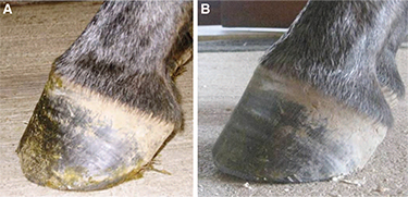 Fig. 3. |
| Fig.3. Amild flexural deformity before trimming(A) and after trimming(B). Note the change in break over created under the dorsal hoof in B |
Treatment
Conservative treatment such as restricting exercise to reduce further trauma is paramount. Correcting the nutritional status of the foal (i.e., weaning the foal to avoid possible excessive nutrition from the mare and/or decreasing carbohydrates), administering an anti-inflammatory agent (NSAID) to relieve pain, administering oxytetracycline to facilitate muscle relaxation, and carefully trimming the hoof are, in the author's opinion, a good starting point. The NSAIDs should be administered short-term and judiciously in foals due to the potential side effects, such as gastroduodenal irritation and nephrotoxicity. For analgesia, the author will administer firocoxib (0.1 mg/kg bwt q 24 hr) or flunixin meglumine (1.1 mg/kg bwt q 24 hr) combined with a gastric protectant. Hoof trimming is directed toward improving the hoof angle by lightly trimming the heels from the middle of the foot palmarly until the hoof wall at the heels and the frog are on the same plane. The bars can be thinned or removed to possibly improve heel expansion, and the heels adjacent to the sulci should be angled to 45° to promote spreading. Breakover is moved palmarly by creating a mild bevel with a rasp, which begins just dorsal to the apex of the frog and extends to the perimeter of the dorsal aspect of the hoof wall (Fig. 3A and 3B). If improvement is noted, this trimming regimen is optimally performed at 2 week intervals. If the toe is constantly being bruised or undergoing abscessation, a hoof compositea,b can be applied to the dorsal aspect of the sole and the distal dorsal aspect of the hoof wall to form a toe "cap" to provide protection. The acrylic composite-impregnated fiberglass or urethane composite used to form the toe cap covers the solar surface to the apex of the frog, protecting that area from further damage. A bevel toward the toe can be created in the composite with a rasp or Dremel© tool to facilitate breakover. If there is adequate integrity of the dorsal section of the hoof wall, the author believes that application of a toe extension to be unwarranted and actually contraindicated. Farriers have traditionally applied toe extensions to create a lever arm using a shoe or a composite, but they only exacerbate wall separation and delay breakover. Furthermore, extensions may contribute to lameness due to excessive stresses on the DDFT when the foal puts full weight on its foot. The above treatment can be temporary, appears to work best when initiated at the first sign of foot deformity before a marked flexural deformity is noted and, when possible, following elimination of any possible inciting causes. The farriery should always be combined with restricted exercise. If the affected foot continues to improve or does not digress, conservative treatment is continued. If a mild flexural deformity progresses in severity to the stage where a marked radiographic flexural deformity is noted, the foal becomes a surgical candidate.
6. Severe Acquired Flexural Deformities (Club Foot)
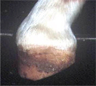 Fig. 4. |
| Fig. 4. Severe flexural deformity. Note the prominent coronet, the steep angle of the dorsal hoof wall, the load being placed on the toe, and the heels of the hoof capsule off the ground. |
The diagnosis is straightforward and based on the characteristic foot and limb conformation described above. Radiographs should be used to confirm the diagnosis and assess changes in the joint. The author will administer mild sedation (half the recommended dose of xylazine [0.33-0.44 mg/kg, IV] combined with butorphanol [0.022-0.066 mg/kg] IV) and place each of the foal's feet on separate wooden blocks of equal height, which allows normal loading of both forefeet. Lateral-to-medial weight bearing images of both forefeet should be obtained. The degree of flexion of the DIPJ, the angle of the dorsal hoof wall, and abnormalities at the margin of the distal phalanx should be assessed (Fig. 5).
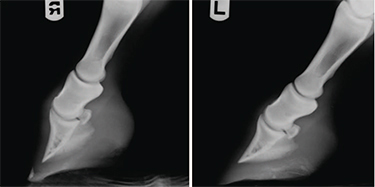 Fig. 5. |
| Fig. 5. Radiographic view of the right front (RF) foot shows a broken forward hoof-pastern axis when compared to the left front (LF) foot which shows normal alignment of the digit. |
Treatment
When a marked flexural deformity is present and confirmed by radiographic examination of the feet, conservative treatment and hoof trimming alone are generally unsuccessful in resolving the problem. Elevating the heels has been advocated to reduce tension in the DDFT and to promote weight-bearing on the palmar section of the hoof. However, although elevating the heels improves the hoof-pastern axis and makes the foal more comfortable initially, the author has not been able to subsequently lower the heel or remove the wedge and establish a normal hoof angle with the heel on the ground. Once a marked flexural deformity of the DIPJ and distortion of the hoof capsule is apparent or progressing, the author recommends transection of the AL-DDFT combined with the appropriate farriery.
7. Desmotomy of the AL-DDFT
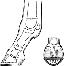 Fig. 6. |
| Fig. 6. Illustration of the lateral side of the foot shows the placement of a reverse wedge after the foot has been trimmed. Illustration of the ground surface of the foot shows the composite wedge reinforced with an aluminum plate. |
8. Aftercare
The surgical aftercare is at the discretion of the attending clinician. Controlled exercise in the form of daily walking or turn-out in a small paddock with firm footing such as a round pen is essential. There is the potential for pain with the initiation of exercise, requiring close monitoring of the foal, and exercise should be increased sequentially. The foal is trimmed at roughly 2 week intervals, based on the amount of hoof growth at the heels with the objective of establishing normal hoof capsule conformation. The composite wedge is removed 1 month after the surgery. At subsequent trimmings, the heels are lowered as necessary from the middle of the foot palmarly, and hoof wall at the toe is trimmed from the dorsal aspect of the hoof wall until the desired conformation is attained. No sole dorsal to the frog is removed. When the desired conformation is reached, the foot is trimmed in a routine manner on a monthly basis. It is important to emphasize that when the hoof capsule returns to an acceptable conformation, only that portion of the sole that is shedding should be removed. This avoids causing discomfort in the dorsal solar area that can result in the horse redeveloping, to some degree, the original deformity.
9. Flexural Deformities in the Mature Horse
Club Feet
 Fig. 7. |
| Fig. 7. A grade 3 club foot on a mature horse. Note the broken forward hoof-pastern axis, the concavity in the dorsal hoof wall, the disparity in hoof wall growth between the toe and the heel, and the poor hoof wall consistency distally. |
The Hoof Capsule Distortion
To apply the appropriate farriery, understanding the proposed mechanism leading to the club foot conformation is helpful. When a flexural deformity is present, the musculotendinous unit is shortened, the degree of which is dependent on the amount of flexion in the DIPJ. This causes a disparity of hoof wall growth, with more growth at the heel than at the toe to compensate for the decreased length of the musculotendinous structures. The frog generally recedes below the hoof wall due to the excessive hoof wall grow that the heels so that the energy of impact is assumed entirely by the hoof wall, bypassing the soft tissue structures and transferring the load directly onto the bones of digit through the laminar interface. The flexural deformity, combined with the excess hoof wall growth at the heels, places the DIPJ in flexion and the distal phalanx in an abnormal alignment relative to the digit; this promotes toe-first landing and, therefore, excessive load on the dorsal section of the joint and hoof capsule. Hoof abnormalities associated with a club foot conformation are thin flat soles, poor hoof wall consistency, especially at the toe, toe cracks, hoof wall separation, and "white line disease".22 Injuries associated with a high hoof angle are thought to include inflammation of the DIPJ due to abnormal loading of the joint, sole bruising, and increased strain on the suspensory ligaments of the navicular bone.19,23
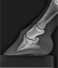 Fig. 8. |
| Fig. 8. Radiograph shows a moderate flexural deformity involving the DIPJ in a horse with a club foot. Note the ideal soft tissue parameters of the hoof capsule that can be assessed on this radiograph. |
Good quality radiographs, consisting of lateral to medial and a weight bearing (horizontal 0°) dorsopalmar views, are necessary for the clinician and farrier to evaluate the condition and apply the appropriate farriery for the club foot. Good soft-tissue detail allows distortion of the hoof capsule to be accessed.9 A lateral to medial radiograph reveals the weight-bearing properties of the foot, allows assessment of the hoof capsule, the position of the distal phalanx within the hoof capsule, solar depth, length of the heels, the osseous integrity of the perimeter of the distal phalanx, and the severity of the flexural deformity of the DIPJ. The degree of flexion indicates the amount of shortening of the musculotendinous unit. The radiographs are used to diagnose any pathology present, determine treatment options, and can be used as a template for farriery (Fig. 8).
10. Therapeutic Farriery
Therapeutic farriery forms the mainstay of treatment for club feet. Farriery should be based on principles rather than a particular method, and the principles remain the same regardless of the severity of the flexural deformity.18,19,22,24 The principles are to achieve normal alignment between the first, middle, and distal phalanges and thus normal orientation and loading of the distal phalanx relative to the ground. Trimming and shoeing is aimed at removing weight-bearing from the toe and dorsal aspect of the distal phalanx and reestablishing weight-bearing to the entire solar surface of the distal phalanx and the corresponding hoof capsule. Historically, farriers have been taught to trim (lower) the heels to correct the distorted hoof capsule and promote weight-bearing in the heel area, but this type of trimming comes with a price. As the severity of the flexural deformity increases, so too does the shortening of the musculotendinous unit; therefore, lowering the heels directly increases the tension within the musculotendinous unit, and these stresses may lead to irresolvable tearing of the dorsal lamellae, widening of the sole-wall junction similar to that seen in the chronic laminitic hoof, and increased pain.18,19,25 The increased forces placed on the DDFT from this type of trimming also promote hoof capsule distortion and abnormal loading. Furthermore, if there is pathology present in the soft tissue structures of the palmar foot, decreasing the height of the heels is likely to place more strain in this section of the foot.
11. Farriery
Distinguishing between a foot with steep hoof angle and a true club foot is important. High hoof angles without phalangeal misalignment or with mild phalangeal misalignment can generally be managed by adhering to good farriery guidelines for trimming such as using the hoof-pastern axis, the center of rotation, and trimming the heels of the hoof capsule to include the frog. It may be necessary with a high hoof angle or mild phalangeal alignment to gradually trim the heels in a tapered fashion from the apex of the frog to the heels. This increases the ground surface of the foot and attempts to reestablish weight-bearing on the entire solar surface of the foot. Breakover is moved palmarly at the same time to compensate for any increased tension in the DDFT created by lowering the heels. This can be accomplished by rolling, rockering, or grinding breakover into the toe of the shoe. If improvement is noted, the horse should be trimmed/shod at 4 week intervals.
Farriery to correct a high hoof angle accompanied by a flexural deformity (club foot) becomes more of a challenge. Again, the object of farriery is to load the heels, compensate for the shortening of the DDFT, and improve the hoof-pastern axis. To accomplish these objectives, farriery is directed at trimming the heels of the hoof capsule, but the amount of heel to remove can be difficult to determine. In mild to moderate club feet, an estimate of how much heel to remove can be made by placing the thick end of a 2° or 3° pad under the toe of the foot and allowing the horse to stand on it 10,19 (Fig. 9). If the horse does not resent the tension this places on the DDFT, this test allows the farrier to safely trim the hoof wall at the heels in a tapered fashion starting in a palmar direction from the widest part of the foot using the thickness of the degree pad as a guide. The toe is shortened by trimming the outer surface of the dorsal hoof wall with a rasp. The trimmed foot is fitted with a shoe that has the break over forged or ground into it starting just dorsal to the apex of the frog and tapering toward the toe to further decrease the stresses on the DDFT. There are also commercial shoes available that have a rockered toe that provide appropriate breakover.
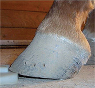 Fig. 9. |
| Fig. 9. A wedge pad can be placed under the toe of a horse with a club foot, which will test the response when tension is exerted on the DDFT. The farrier can then determine the amount of hoof wall that can be safely removed from the heels of the hoof capsule. |
With the more advanced cases of club feet, the heels should still be lowered to load the heels and unload the toe, but the addition of heel elevation following the trim is necessary to compensate for the shortening of the musculotendinous unit. The concept of lowering the heels with the trim then wedging the palmar aspect of the hoof back up is often not understood. When the heels are trimmed back to the widest point of the frog, the load bearing surface area of the foot increases, and this is necessary for normal function of the hoof. However, the musculotendinous unit must be accommodated and maintained without excessive tension and pain. This is accomplished by decreasing the breakover and by adding elevation to the palmar aspect of the hoof. The degree of wedge that is applied often mimics the amount of heel removed, but in many cases may be less due to mechanical contributions made by rockering or rolling the toe of the shoe. The amount of heel elevation needed if necessary can be demonstrated following the trim by placing the trimmed foot on the ground 6 to 8 inches palmar to the contralateral limb. A space will generally appear between the heels of the foot and the ground (Fig. 10). The author uses either a wedge shoe or places a degree pad or a bar wedge between the heels of the foot and the shoe to compensate for the shortening of the muscle-tendon unit (Fig. 11). This method allows the heels to be weight-bearing but at the same time decreases the stresses on the musculotendinous unit. Creating breakover in the shoe to further relieve stress in the DDFT, as described above, is essential. It is important to note that when the heels are elevated with a wedge shoe, the normal ground reaction forces and load bearing structures are altered. To redistribute the load, it is beneficial to apply a "pour-in" pad or impression material to the sole of the hoof between the branches of the shoe to reestablish load sharing of the weight bearing structures of the hoof (Fig. 12). Without increasing the surface area over which the ground reaction force is distributed, the heels may become overloaded over time and possibly result in quarter cracks, contracted heels, and subsolar bruising of the heel region. Following the farriery, it is necessary to see the horse have a flat strike pattern rather than a toe first landing. If the horse does not land flat, heel elevation should be considered.
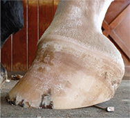 Fig. 10. |
| Fig. 10. Post trimming, the foot is placed 6 to 8 inches palmar to the contralateral limb. A space under the heels indicates shortening of the musculotendinous unit and the necessity of heel elevation. |
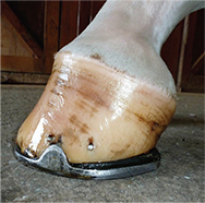 Fig. 11. |
| Fig. 11. Club foot illustrated in Fig. 7. Following the appropriate trim, the horse is shod with a wide web steel shoe with heel elevation and breakover created in the shoe. |
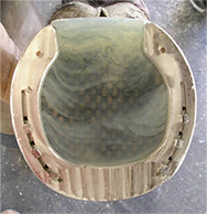 Fig. 12. |
| Fig. 12. Pour-in pad used to change the distribution of pressure between the branches of a shoe with wedge heels. |
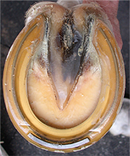 Fig. 13. |
| Fig. 13. Polyurethane shoe attached directly to the foot with an acrylic adhesive (photo courtesy of Curtis Burns). |
In severe cases of club feet, the hoof wall consistency may be compromised such that alternative methods of application (other than nailing) may need to be integrated. The most common method of "gluing" a shoe on is termed "direct gluing," which attaches a shoe directly to the weight bearing surface of the hoof wall and sole via some type of adhesive. Aluminum rather than steel shoes are typically used for this purpose because aluminum is very porous and the adhesives adhere with more affinity. The same principles for shoe fit are used with glue-on shoes vs. shoes that are nailed. Recently, polyurethane shoes have reached the marketplace and can be applied via the direct gluing technique (Fig. 13). These shoes are mentioned because they are flexible, allow the heels to expand, yet still carry the advantage of being attached with an adhesive. Although glue-on shoes will not take the place of traditional farriery, they provide an alternative when necessary to prevent the compromised club foot from continuing to lose sole depth and develop further hoof capsule distortion by not having a shoe applied. It is always the goal, however, to transfer the horse into the most normal and simple shoeing/trimming protocol when possible.
12. Farriery Combined With Surgery
In selected cases, horses with a severe flexural deformity or horses that have not responded to appropriate farriery and remain lame may benefit from a desmotomy of the AL-DDFT.2,10,18,19,21,23,25 This release procedure, along with therapeutic farriery, allows realignment of the distal phalanx within the remainder of the digit, loads the entire surface of the hoof capsule, and readily allows the accompanying distortion of the hoof capsule to be improved. It is the author's opinion that if this surgery is being contemplated, it should be performed early in the horse's athletic career, before there is a significant hoof capsule distortion and before pathological changes involving the DIPJ and or the margin of the distal phalanx become evident on radiographs.
The surgery is generally performed under general anesthesia but in the mature horse, it can be performed standing using sedation and local or regional analgesia if necessary. If surgery is performed with the horse standing, the heel should be elevated by taping a 12° wedge to the foot to decrease tension in the AL-DDFT/DDFT complex allowing the ligament to be easily identified, separated, and transected away from the cutaneous incision. The client should be forewarned that the surgery involves an extended recovery period, and a blemish or fibrous thickening at the surgery site is inevitable due to the mature nature of the tissue. Caution is advised when performing the farriery that accompanies the surgery because the soft tissue structures within the hoof capsule and the digit have adapted/ accommodated for the distortion of the hoof capsule. The author will trim the palmar section of the foot moderately using the information obtained from the radiograph for guidance and then apply two or three 2° wedge pads using either a shoe or a cuff (Fig. 14). After surgery, the horse is walked daily, and a degree pad is removed every 7 to 10 days depending on the comfort of the horse. After 3 weeks, the horse is allowed turnout in a small paddock such as a round pen for an additional 3 weeks and then turnout in a larger area for 3 to 6 months before exercise is resumed. The appropriate farriery must be maintained post surgery or the foot will have a tendency to revert to the original foot conformation. The cosmetic appearance of the limb is maximized by keeping the limb bandaged for the first 6 weeks. In a limited number of cases that the author has managed or consulted on, the benefits returning the horse to soundness have far outweighed the rehabilitation process being labor intensive. The author has not realized any benefit in applying a toe extension to the shoe in the adult horse following surgery. Many mature horses with a club foot frequently have damage to the dorsal lamellae and a stretched white line similar to that found in horses with laminitis, and therefore, toe extensions may markedly exacerbate detrimental mechanical forces on the lamellae and dorsal hoof wall.
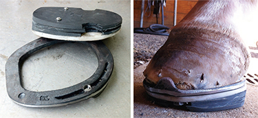 Fig. 14. |
| Fig. 14. Shoe with three removable 2° wedge pads attached to a plastic treatment plate. |