|
Can farriery resolve the negative palmar angle? Stephen E. O’Grady, DVM, MRCVS Reprinted with permission. Original article appeared in The Natural Angle Vol 19: Issue 1, 2022
Introduction The equine hoof can be divided into dorsal and palmar/plantar sections. The deformable soft tissue structures located within the palmar/ plantar section of the hoof capsule are the frog, frog corium, digital cushion, ungual cartilages and the deep digital flexor tendon. The function of these structures is to absorb concussion, dissipate the energy of impact and decrease the vibrations associated with the foot landing on the ground. These soft tissue structures can accept or share some weight-bearing function but unfortunately, they are often subject to compromise when excessive load is placed on this section of the foot over time. This deterioration in the palmar foot structures is not readily apparent when the horse is barefoot (unless previously shod), so there must be a correlation to the application of shoes. However, there appear to be multiple factors that may contribute to this demise, such as:
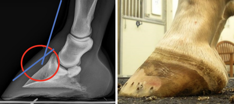
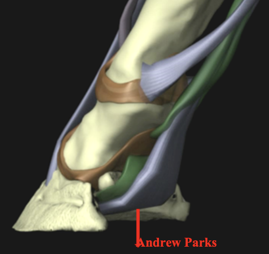
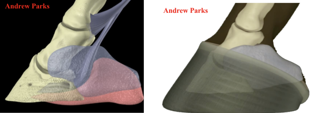
Damage to the integrity of the soft tissue structures and a loss of structural mass result in varying degrees of a negative palmer/plantar angle. The negative palmar angle (NPA) or more correctly, ‘the angle of the solar border of the distal phalanx’, has become a diagnosis, a disease, and a focus for a myriad of veterinary / farriery treatments. Simply stated, the negative palmar angle results from a loss of the soft tissue structures (especially the digital cushion) in the palmar section of the foot. The loss of mass allows the distal phalanx to descend distally in the palmar section of the hoof capsule. The result is a change in the position of the distal interphalangeal (DIP) joint which places the joint in dorsiflexion and leads to a distortion of the hoof conformation. This is not to say that the NPA does not or cannot also have negative consequences on soundness...it does! The change of position of the bone creates a broken back hoof-pastern axis (HPA), changes the position of the (DIP) joint which changes the distribution of weight bearing on the solar surface of the foot and increases the tension in the deep digital flexor tendon (DDFT) (Figure 1). The decreased mass of the soft tissue structures in the palmar foot hinders the foot from dissipating the energy of impact and providing adequate shock absorption. Many are not aware of the sling formed by the DDFT under the navicular bone that descends into the soft tissue structures in the palmar foot as the joint moves in a distopalmar direction when the heel strikes the ground, thus forming one of the initial shock absorbing mechanisms of the foot. This primary shock absorbing mechanism is lost when a significant decrease in soft tissue mass occurs (Figure 2). All these changes can have deleterious effects on the equine foot and soundness because the functionality of the palmar foot is lost.
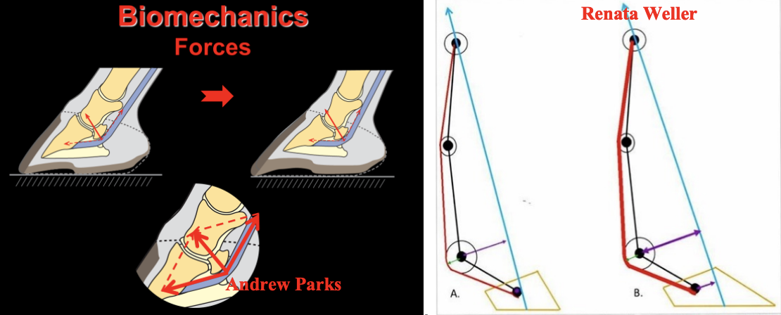
The biomechanical aspects and the correlation to lameness related to the effect of the negative angle of the distal phalanx on the podotrochlear apparatus (navicular area) is huge. Eliashar (Eliashar et al EVJ 2004) showed that a 1° decrease in palmar angle of the distal phalanx results in a 4% increase in peak force on the navicular bone. This places excessive load on the navicular bone, the navicular bursa, and the distal extent of the DDFT (Figure 3). Furthermore, the horse will now spend a substantial amount of time with the DIP joint in dorsiflexion which is not only an abnormal position of the joint but also changes the pattern of weight bearing on the solar surface of the foot.
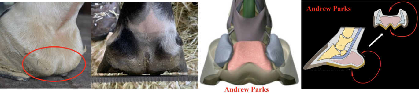
Improving hoof conformation to address the negative palmar angle of the distal phalanx presents clinicians with a farriery challenge. Often improvement cannot be achieved and’ therefore, the focus is to maintain the compromised structures. Traditional farriery methods used are to provide heel elevation to improve the digital alignment and reposition the DIP joint. However, this practice can be detrimental if the soft tissue structures in the palmar foot are markedly compromised or lack the appropriate mass. When heel elevation is added, it temporarily improves the appearance of the hoof capsule and the HPA, but the pressure placed on the soft tissue structures overtime will damage them further. The limiting factor when attempting to improve the structures in the palmar foot is the inability to grow hoof wall in the heel area necessary to improve the hoof capsule. The primary soft tissue structure involved in the compromised palmar foot is the digital cushion. Genetics are incriminated for the decreased size of the digital cushion as many animals are born (TBs for example) with inadequate tissue mass in the palmar foot while other factors, in the author’s opinion, are lack of foot development as a juvenile, shoes applied too early, training regimens, overuse, surfaces and inappropriate farriery practices. Considering the type of cells that comprise the digital cushion, there is little evidence to suggest that it can be restored or increase in mass. When the mass of the digital cushion is decreased, more weight is placed on the frog which causes it to enlarge and often protrude or prolapse below the solar surface of the foot. It is the author’s opinion, based on many years of farriery, that hoof wall growth requires a structural framework between the heels of the hoof capsule to produce horn. Loss of that framework occurs when the mass of the soft tissue structures in the palmar foot is markedly decreased (Figure 3).
Farriery Various farriery options are used to address this problem: heart bar shoes, roller motion shoes, frog support pads, heel plate shoes, stabilizer (spider) plates, etc....all with varying degrees of success and usually related to the amount of compromise in the soft tissue structures. There is a symbiotic relationship between the osseous structures in the dorsal section of the foot and the soft tissue structures in the palmar/plantar foot. For a healthy foot, the importance of this interrelationship cannot be over emphasized in maintaining structural integrity and proper intracapsular location of anatomic components (Figure 4). The NPA conformation creates a puzzle for the farrier but by using the pieces representing the anatomy, biomechanics and basic farriery principles, an overall picture of the problematic hoof capsule can be generated, and a farriery plan formulated. Appropriate farriery for this condition requires careful evaluation of the foot conformation, assessing the amount of damage to the structures of the palmar foot, the landing pattern of the horse, the weight of the horse, the surface on which the horse works and the proper farriery necessary to address the amount of compromise. Farriery must be based on principles such as the appropriate trim, protection of the compromised structures, redistribution of forces and placement of all palmar foot structures in a ‘load sharing’ position. The trim is always the initial step, especially to improve hoof conformation/distortions. The type, size and placement of the shoe are intended to protect the trim, increase surface area, and add the necessary biomechanics. Finally, there are many farrier products and synthetic materials available; that, when combined with the appropriate trim and shoe, can add resiliency to improve the function of the palmar foot. There is no single answer, method or ‘one size fits all’ that can be used for consistent success when addressing the NPA.
Summary The NPA poses a constant dilemma to both the veterinary and farriery professions with regards to a healthy foot and continuing soundness. The position of and the forces on the distal interphalangeal joint resulting from the NPA can be a source of pain and one of the most common joints treated in veterinary medicine. However, any medication used to treat this joint will only have transient benefits unless the conformation of the palmar foot is addressed and improved. Another example that a good veterinarian farrier relationship compliments both professions and ultimately benefits the horse. |