Low or under run heels in the hind feet.Reprinted with permission from the Farrier Products Distribution. |
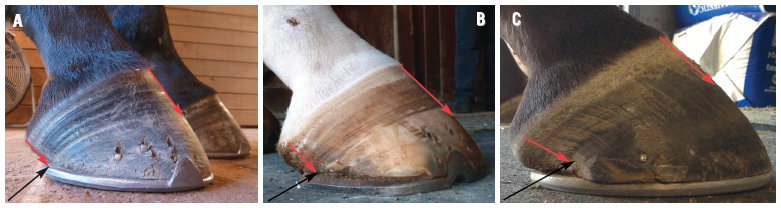 |
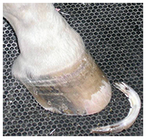
| Figure 1. Hind feet with a low heel - A is the early stage, B is more advanced and C is severe. Note the red arrows showing the disparity in the growth rings and the 'bull nose' forming. The black arrow denotes the end of the heel. |
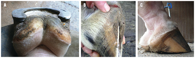 |
| Figure 2. Figure 2A shows the position of the frog prolapsed between the branches of the shoe. Note the frog positioned below the hoof wall when the shoe is removed (Fig 2B) and the horse standing on the frog when the foot is on the ground (Fig 2C). |
 |
| Figure 3. Note the 'trough' present between the apex of the frog and the inner margin of the shoe. |
CLINICAL EXAMINATION OF THE FOOT
Abnormal conformation of the hind feet is easy to recognize. When looking at the limb from the side, the digit will show a broken back hoof-pastern axis. The slope of the coronary band from the toe to the heel will have an acute angle at the heel and the bulbs of the heels will have a 'knob' shaped appearance which can be seen lying against the shoe. There will be a disparity in the growth rings below the coronet from the toe to the heel and the dorsal hoof wall will assume a "bull nosed" appearance (Figure 1). Looking at the foot from behind, the frog is often situated well below the hoof wall and the frog can be seen to prolapse down between the two branches of the shoe (Figure 2). The shape of the frog is generally large from the constant stimulation with the ground. Upon removing the shoe, the end of the heel of the hoof wall is located well forward from the base of the frog and the horn tubules will be parallel with the ground. The hoof wall at the heel will be thin, there will be no angle of the sole, and the bars may be absent. When the foot is placed on the ground, total weight bearing will be placed on the frog and many horses are reluctant to stand on the frog when the opposing limb is lifted off the ground. Viewing the toe area on the ground surface of the foot, there will be a large "trough" noted between the apex of the frog and the inner branch of the shoe instead of a smooth transition of the sole from the frog to the sole wall junction (Figure 3). Hoof testers placed on either side of the heel at the angle of the sole will often elicit a painful response and the structures will deform.
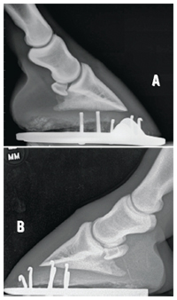 |
| Figure 4. Figure 4A is moderate low heel conformation while figure 4B is severe. Note the position of P2 relative to P3 on both radiographs. This places the load on the palmar section of the foot. Also note the 'knob' appearance at the heel bulb. |
RADIOGRAPHS
A lateral radiograph of the hind foot will show a broken back hoof pastern axis with the middle phalanx (P2) being pushed plantar and distally relative to the distal phalanx (P3) during weight bearing (Figure 4). This places excessive stresses on the plantar section of the joint capsule. The solar margin (plantar angle) of the distal phalanx is lower than the dorsal margin of the distal phalanx. The sole depth below the dorsal margin of the distal phalanx is markedly increased relative to the sole depth at the heel and the perimeter of the distal phalanx can be seen migrating toward the dorsal hoof wall. This is what causes the "bull nose" appearance of the dorsal hoof wall. The soft tissue structures in the plantar section are noted to be lying against the shoe.
FARRIERY
Damage to the heels of the hind feet is easier to improve than in the forefeet, possibly due to the anatomy and the difference of the load encountered on the hind limbs. The severity or the distortion of the hind feet will obviously be proportional to the amount of time the condition has been present and thus harder to resolve. The first part of the farriery is to address the frog being located below the hoof wall. If severe, the horse should be taken out of work and allowed to go without hind shoes briefly (as little as 5-7 days may be all that is necessary). The shoes are removed and the hoof wall is trimmed from quarter to quarter according to the sole depth (Figure 5). The horse is then kept on a firm surface, thus placing pressure on the frog which quickly assumes the same plane as the heels on either side (Figure 5). If the horse needs to continue in work and wear shoes, the approach can be modified. The hind shoes are removed a day or two before the horse is due to be shod and the foot is trimmed as described above. A degree pad is cut out to fit the foot and secured to the foot with brown gauze and elastic tape. The horse is placed in a stall on a firm surface for 24-48 hours.
 | 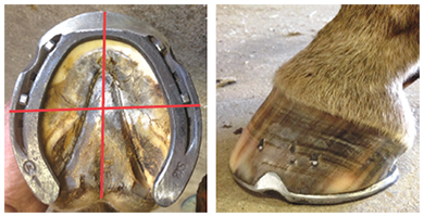 |
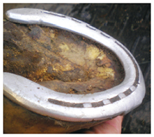
| Figure 5. Excess hoof wall is removed from toe quarter to toe quarter. This will decrease sole depth at the toe and improve the angle of the solar border of the distal phalanx with the ground. | Figure 6. Ground surface and side view of a hind foot shod with a Kerckhaert SX-8 shoe. Red lines show the placement of the shoe and the dimensions of the foot being as wide as it is long. Note the smooth transition of the sole from the apex of the frog dorsally. Also note the breakover created in the shoe. Side view shows the placement of the shoe and the shoe extending marginally beyond the heels of the hoof capsule (Courtesy of Jeff Ridley, CJF). |
When the wedge pad is removed, the frog and the hoof wall will be on the same plane, forming a flat even surface which includes the frog and both heels. The horse is then shod paying strict attention to the trim. Any additional horn at the heels can be removed so the heels of the hoof wall are solid and approach the base of the frog, being careful to keep the frog and both heels in the same plane. When the hoof wall and the frog are on the same plane, the load is shared across the plantar section of the foot. A shoe can now be fitted and applied. We fit shoes to the hind feet the same as the front where a line is draw across the widest part of the foot and the shoe is fitted so the line is placed in the middle of the shoe3. In the hind feet, the branches of the shoe may extend marginally beyond the end of the heels (Figure 6). Breakover should be created in the hind shoe either by forging or using a hand grinder beginning at the inner margin of the shoe and tapering toward the periphery of the shoe. Placing breakover in the shoe should be no different than the front feet as the biomechanics remain the same. A recent paper in the literature showed a smoother shift in the center of pressure and a more fluent hoof enrollment using a rolled toe shoe on the hind feet4. In order to keep the frog and hoof wall on the same plane or if mild heel elevation is necessary, a metal or aluminum heel plate or a leather wedge can be placed under the shoe at the heels as long as the shoe is fitted in the manner described above. This will concentrate the load across the frog and heels rather than behind the heels which is the case with a long shoe or trailers. This is usually a temporary measure and can be discontinued once the heels have stabilized.
| GLOSSARY OF FARRIER TERMS USED IN THIS ARTICLE Anatomical termPhalanx Common nameBone Description Any of the principal bones of the digit. Anatomical term Distal Phalanx Common nameCoffin Bone, P3, Pedal bone Description Bone enclosed within hoof capsule. Anatomical term Distal Interphalangeal Joint (DIP) Common nameCoffin Joint Description The joint between the middle(DIP) and the distal (P3) phalanx which includes the navicular bone. Anatomical term Plantar Common nameDirection Description The caudal facing aspect of the hind limb from the tarsus (hock) distally (toward the ground). |
CONCLUSIONS
Damage to the heels of the hind feet is much easier to resolve or improve than in the fore feet. This could be due to the anatomy of the hind limb along with the shape and function of the hind feet. Once the frog has been repositioned and the heel structures have grown, attention to the foot prep is necessary to keep the frog and heels of the hoof wall in the same plane. The trim, along with the size and placement of the shoe, are equally important in maintaining the health of the heels of the hind feet.