1. Introduction The hoof capsule is comprised of the hoof wall, sole, frog, and bulbs of the heels, which, through the unique continuous bond between its components, form a casing on the ground surface of the limb that affords protection to the soft tissue and osseous structures enclosed within the capsule.
1 The hoof wall is a viscoelastic structure that has the ability to deform under load and then return to its original shape when the weight is removed. It is well accepted that abnormal weight distribution on the foot or disproportionate forces placed on a section of the hoof will, over time, cause it to assume an abnormal shape.
1-4 These abnormal stresses within the foot will also predispose the foot to injury or disease. Increased stress or weight-bearing placed on a section of the hoof capsule may originate from a single source or it may be from multiple contributing factors such as abnormal limb conformation, strike pattern, amount of work, type of footing, and inappropriate farrier practices. Excess stress placed on one section of the hoof capsule can manifest itself in a variety of ways, such as compressed growth rings, flares or under-running of the hoof wall, dorsal migration of the heels, and either focal or diffuse displacement of the coronary band.6,7 Distortion of the hoof capsule of the forelimbs appears to be related to limb alignment and load, whereas deformation in the hind feet appears to be different and related to propulsion. Because the hoof capsule distortion of the forelimbs is commonly associated with lameness and various disease processes, only the forelimbs will be considered in this report. Because the "normal" foot has never been defined, each view will begin with what is perceived to be an ideal, good, or healthy foot.
1,8 Palpation of the hoof capsule often complements the visual examination, and the areas where palpation is relevant will be included. The goal of evaluating the hoof capsule is to identify deformation and changes in growth pattern that indicate abnormal distribution of forces (stresses) on the foot. Because hoof capsule distortion and abnormal loading usually accompany lameness, farriery will form part of or sometimes the entire treatment. Farriery is used to help redistribute the load and help improve or resolve the hoof capsule deformation.
2. Mechanism of Distortion Evaluation of the hoof capsule morphology will indicate where the hoof wall is unduly stressed; however, the evaluation must be coupled with an understanding of the abnormal distribution of forces that lead to hoof capsule deformation. Understanding the biomechanical forces leading to hoof capsule distortions is also helpful for the clinician in applying the appropriate farriery to modify these stresses. There are many excellent reviews of basic biomechanics of the hoof in the veterinary literature. 1-5 Increased load or weight-bearing by a portion of the wall has three consequences: (1) it may cause deviation of the wall outward (flares) or inward (under-running) from its normal position; (2) it may cause the wall to move proximally; or (3) it may decrease hoof wall growth. A reduction in load or weight-bearing generally has the opposite effect. Briefly, in the standing horse, the weight of the horse borne by the limb is supported by the ground, which opposes the weight with an equal and opposite force. The force exerted on the foot by the ground is termed the ground reaction force. The term center of pressure (COP) is the point on the ground surface of the foot through which the ground reaction force acts on the foot. The center of pressure varies among horses but is approximately located in the center of the solar surface of the foot in the standing horse. However, when the horse is moving, the location of the COP changes dynamically. The position of the COP at any point in the stride determines the distribution of forces between the medial and lateral and the dorsal palmar aspects of the foot. When the center of pressure is moved to one side of the foot, that side of the foot will be subject to increased forces. If the COP is moved in a palmar direction, the weight-bearing or load on the palmar hoof wall is increased. Relating this to hoof capsule distortions, if the COP is located more medially, over time, a medial hoof wall flare (bending) and a lateral under-running will develop. Or, if the COP is located more dorsally because of increased tension in the deep digital flexor tendon, the hoof capsule will develop a higher heel with a flare in the dorsal hoof wall. Farriery is used to change the location of the center of pressure (to some extent) and change the distribution of forces on the ground surface of the foot.
3. Limb Conformation When evaluating hoof capsule deformation, limb conformation should be considered. Abnormal limb conformation affects the landing pattern and stance phase of the stride. Few horses have ideal limb conformation, and any change in conformation is going to change the distribution of forces within the hoof capsule, leading to deformation. In the frontal plane, the forelimbs should be of equal length and size and bear equal weight. A line dropped from the scapulohumeral joint to the ground should bisect the limb. Certain types of abnormal limb conformation have been described.9 In the frontal plane, abnormal conformation is described as valgus (the limb's segment distal to the affected joint will deviate laterally) or varus (the distal segment of the limb will deviate medially). The joint most often affected is the carpus, and, to a lesser degree, the metacarpalphalangeal joint. Here, there will be excess load placed on the hoof opposite the direction of the deviation. If a line dropped from the metacarpalphalangeal joint through the digit to the ground does not bisect the hoof capsule, the foot is considered offset to one side (usually laterally) and therefore increased load is placed on the opposite side of the foot (Fig. 1).
| |
| Fig. 1. Distal phalanx within hoof capsule will be offset laterally. Coronary band will be displaced proximally on media quarter/ heel. |
In the transverse plane, conformation abnormalities are characterized by axial rotations of the limb or its segments, either laterally or medially. For example, a horse with a narrow chest and a lateral axial rotation will land on the lateral side of the hoof and then load the medial side resulting in proximal displacement of the quarter /heel on the medial side and causing the hoof deformation termed "sheared heels"10,11 (Fig. 2). A limb with a medial (inward) rotation of the digit relative to the third metacarpal bone (toed-in) will develop a hoof with a diagonal asymmetry, with a narrow lateral toe and medial heel and a wide medial toe and lateral heel. The altered distribution of forces leading to hoof capsule deformations follow a logical pattern in which the overloaded sections of the hoof are less developed and the under-loaded sections are overdeveloped. In the sagittal plane, abnormal conformation can best be described by the position of the distal interphalangeal joint (DIP), either a flexural
| |
| Fig. 2. Horse with one heel bulb displaced proximally (sheared heel conformation). Note the contour of the pastern above the displaced heel bulb. |
deformity or marked dorsiflexion (ie, extension) of the joint. The shape or conformation of the hoof in the sagittal plane will be dependent on the tension in the deep digital flexor tendon, the integrity of the laminar apparatus, and the digital cushion-all of which determine the angle of the solar margin of the distal phalanx. A flexural deformity will overload the toe, whereas marked dorsiflexion of the DIP joint will overload the palmar section of the foot.
4. Evaluation of the Hoof Capsule A detailed morphological examination of the foot should begin with observing the horse in motion, both going away from and toward the examiner, on a firm, flat surface to note the landing pattern. The foot is then viewed from all sides while it is on the ground. Finally, the ground surface is examined with the foot off the ground. Additionally, small changes in the shape of the hoof capsule (such as the coronet and the digital cushion) may be better appreciated by careful palpation of the foot than by visual inspection.
| |
| Fig. 3. Dorsal view: the hoof should be symmetrical. A line drawn between any two comparable points on the coronary band should be parallel to the ground. The hoof should be symmetrically related to the distal limb such that a vertical line should bisect the third metacarpal bone, the pastern, and the hoof (courtesy of Dr Andy Parks). |
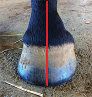
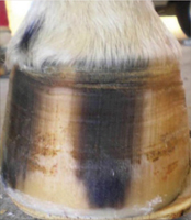
5. Dorsal Aspect When the foot is viewed from the dorsal aspect, the ideal hoof should be approximately symmetrical. An imaginary line drawn between any two comparable points on the coronary band should be parallel to the ground. The medial wall should be the same height as the lateral wall, but because it is often slightly steeper, it may be slightly shorter. An imaginary line that bisects the third metacarpal should bisect a line drawn between any two comparable points on the coronary band or the ground surface of the hoof. Similarly, the hoof should be symmetrically related to the distal limb such that an imaginary line that bisects the third metacarpal bone, bisects the pastern and the hoof, allowing for the slight asymmetry caused by the different angles of the medial and lateral wall (Fig. 3).1 When the foot is viewed from the dorsal aspect, the shape of the forefeet may be asymmetrical, with one hoof being narrower than the other ("mis-matched feet"). Several abnormalities may be visible at the toe/ quarters such as flares or under-running of the wall. The coronary band may be unevenly distributed, most commonly by an uneven slope from one side of the foot to the other. A distally directed arch in the coronary band in the dorsal portion of the foot usually indicates extensive remodeling of the distal phalanx at the toe. Examination of the growth rings below the coronet may show divergence of the rings from one side to the other indicating uneven or excessive load (Fig. 4). The angulation of the dorsal horn tubules toward the medial or lateral side of the hoof capsule should be noted; normally they are
| |
| Fig. 4. Note divulgence of growth rings below the coronet from the lateral to medial side. |
parallel, so when they appear tilted medially or laterally, it suggests that the whole hoof capsule may be tilted in that direction (Fig. 5). The position of where the pastern bisects the hoof capsule should be noted, that is, is its entry on the midline or displaced medially or laterally?
On palpation, the coronary band of a healthy hoof should feel thick and spongy. There should be no evidence of a "ledge" or "trough" behind the proximal margin of the hoof capsule when palpated. A depression in the coronary band indicates that the distal phalanx has displaced within the hoof capsule, a finding that can be present in sound horses.12 This palpable depression will generally be accompanied by a thin, flat sole, narrow frog, and contracted heels. The dorsal aspect of the coronary band should also be palpated for effusion of the DIP joint. This is often seen with horses that have a broken back hoof-pastern axis.
| |
| Fig. 5. Hoof with a separation in the dorsal hoof wall. Note the angulation of the horn tubules toward the medial side of the foot. Also note the focal "arch" in the coronet on the lateral side. |
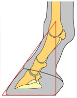
6. Lateral Aspect When viewed from the lateral aspect, the angle the dorsal hoof wall forms with the ground is variable and typically related to the conformation of the digit. The heel tubules of the hoof capsule should form an angle with the weight bearing surface similar to the angle of the horn tubules in the toe region. Tradition has it that the angle of the wall at the heel should match that of the dorsal hoof wall; however, it is usually a few degrees less. As the foot accepts weight, it expands and the ground surface at the heels moves against the shoe, causing wear that decreases the heel angle. The amount of wear is dependent on the integrity of the structures in the heel. The length of the dorsal hoof wall is similarly variable but is determined by the amount of sole depth present. There are two guidelines that relate the proportion of the foot to the rest of the distal limb.
| |
| Fig. 6. Conformation of the foot as it relates to the digit can be depicted with a triangle. A line drawn down the dorsal surface of the pastern and hoof is the hoof-pastern axis. A vertical line that bisects the third metacarpal bone should intersect the ground at the palmar aspect of the heels of the hoof capsule. Connect these two lines with the angle of the dorsal hoof wall and the ground surface of the foot to form a triangle (courtesy of Dr Andy Parks). |
First, the foot-pastern axis describes the relationship between the angles made by the dorsal hoof wall and the dorsal aspect of the pastern with the ground. Ideally, the dorsal hoof wall and the pastern form the same angle with the ground so that the angle between them is 180° and the axis is considered straight. Second, an imaginary line that bisects the third metacarpal should intersect the ground at the most palmar aspect of the ground surface of the hoof. These two guidelines used in conjunction with the angle of the dorsal hoof wall and the ground surface of the foot combine to form a triangle of proportions that represents the relationship between the hoof and the distal limb regardless of the size of the horse (Fig. 6).1 Evaluation of the hoof capsule from the side view should begin with the coronet as this structure can provide very useful information. The healthy coronary band should have a gentle, even slope from the toe to the heels, and the hair should lie flat against the hoof capsule; hair projecting horizontally may indicate excessive forces on the associated hoof wall.
13 The coronary band is dynamic, and its shape can be affected by chronic overloading.
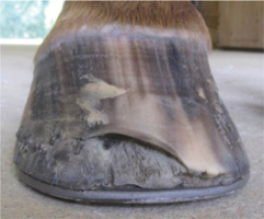
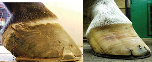
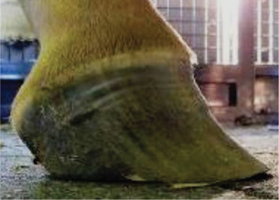
14 A proximally directed diffuse arch at the quarters or a focal directed arch toward the heels is evidence of chronic overloading of that section of the foot (Fig. 7). A coronary band with an acute angle at the heels relative to the ground that bends distally at the heel to form a "knob" appearance is an indication that the foot has poorly developed or under-run heels and the hoof wall at the heels has migrated dorsally (Fig. 8).
| |
| Fig. 7. A section of the coronet in the palmar section of the foot has been displaced proximally (focal arch). Note the relationship of the heel of the shoe and the origin of the defect, which denotes excessive load. The foot on the right shows a change in angulation of the horn tubules, curvature of the growth rings, and a proximal displacement of the coronet (dorsal arch). |
| |
| Fig. 8. Foot with under-run heels showing the "knob" appearance. Note the curve in the growth rings as the heels migrate dorsally. There will often be a depression ("thumbprint") showing the extent the heels of the hoof capsule have migrated forward (arrow). Palmar view shows the decrease in structural mass of the digital cushion. |
| |
| Fig. 9. Clubfoot. Note the coronary band has lost the slope and is almost parallel with the ground. Also note the flare in the dorsal hoof wall. |
A coronary band that is horizontal relative to the ground and often accompanied by a flare in the dorsal hoof wall would denote an upright or clubfoot conformation (Fig. 9). Asymmetry of the height of the coronary band in the quarter/heel region on one side occurs when the horse develops a "sheared heel," a hoof capsule distortion resulting in proximal displacement of one quarter/heel bulb relative to the contralateral side of the foot.
15 The medial heel bulb/quarter is more commonly displaced proximally, as it is more common for the foot to be offset laterally. The angle of the coronary band can be used to estimate the position of the distal phalanx within the hoof capsule. One study described the angle of the coronary band of apparently normal front feet to be 23.5° ± 3°.
16 If the angle of the coronary band is >45°, the plane of the solar margin of the distal phalanx will decrease. At the other extreme, a coronary band parallel to the ground is indicative of a high palmar angle, which is often associated with a club foot or rotation of the distal phalanx. The width of the growth rings below the coronet should be equal from toe to heel. A disparity in the width of the growth rings between the toe and the heels is indicative of non-uniform circulation of the coronary corium or excessive forces below because wall growth is generally inversely related to load. An example of this disparity would be chronic laminitis typified by more horn growth at the heels than toe growth. However, regional irregularity in spacing of growth rings is not uncommon; the most frequently observed is a decrease in spacing at the quarter associated with proximal displacement of the coronary band as noted with sheared heels.10 The angulation of the horn tubules from dorsal to palmar should be noted because horn tubules that are parallel with the ground in the heel area are associated with under-run heels. Flaring or concavity of the dorsal hoof wall accompanied by underrunning of the heels is readily appreciated from the lateral side. The presence of hoof wall flares or cracks are often caused by chronic, excessive overloading of the hoof wall in the region in which these defects are found.
10,11,14,17 Vertical cracks in the quarter are more likely to occur with a sheared heel. Horizontal cracks are usually the result of a disruption of production of horn caused by coronary band trauma or when a subsolar infection ruptures at the coronary band.
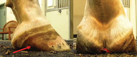
7. Palmar Aspect The heels are evaluated from the palmar aspect for their overall width and height. The heels frequently become narrowed when the foot itself is narrow. Additionally, the central sulcus of the frog may extend proximal to the hairline so that a cleft becomes apparent in the skin of the pastern between the heels. The overall height of the heels is readily assessed from the lateral aspect, but viewing from the palmar aspect is useful to compare the relative heights of the two heels. For example, in the case of the sheared heel, one heel is displaced proximally in relation to the other. Another example is mismatched feet in which there is a marked disparity in heel height. The contour of the junction of the heel bulbs with the skin can be evaluated relative to the width of the hoof wall at the heels and the thickness of the digital cushion (Fig. 10).
| |
| Fig. 10. A, Illustration shows various contours of the junction of the heel bulbs with the skin; B, wide heel; C, contracted heel with a poor digital cushion. Note also the sheared heel on this foot. |
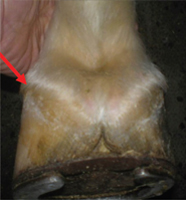
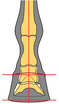
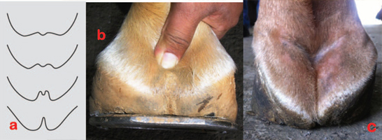
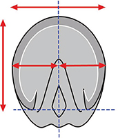
8. Distal or Solar Aspect When viewed from the distal surface, the ground surface of the foot should be approximately as wide as it is long. The foot should be approximately symmetrical about the long axis of the frog; the lateral side of the sole frequently has a slightly greater surface area that corresponds with the difference in wall angles at the quarters described in the dorsal view. The width of the frog should be approximately 60% to 70% of its length. The ground surface of the heels should not project dorsal to the base of the frog. Imaginary lines drawn across the most palmar weight-bearing surface of the heels and across the heel bulbs at the coronary band should be parallel and both lines should be perpendicular to the axis of the frog (Fig. 11).
| |
| Fig. 11. The ground surface of the foot should be approximately as wide as it is long (red lines) and approximately symmetric about the long axis of the frog (blue lines). The heels should not project dorsal to the frog (courtesy of Dr Andy Parks). |
1 If a three-dimensional object such as the foot changes in one plane, it will change in at least one other plane. Therefore, examination of the ground surface of the foot reveals much about the changes in the wall of the hoof capsule. For example, if the contour of the wall is displaced away or toward the median plane in the dorsal two thirds of the foot, this usually corresponds with a flare or under-running of the wall, respectively. If only one heel buttress is displaced dorsally in relation to the base of the frog, it usually corresponds with the proximal displacement of that heel plus or minus the quarter termed sheared heel.
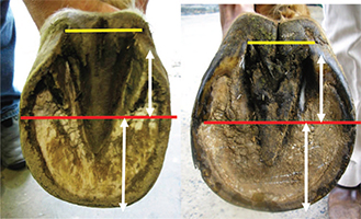
The author begins the evaluation of the solar surface of the hoof capsule by drawing a line across the widest part of the foot. This line forms a consistent landmark and is located just dorsal to the center of rotation (of the distal interphalangeal joint). With the use of this line as a starting point, there should be approximate proportions from this line to the perimeter of the toe and to the base of the frog (Fig. 12).
| |
| Fig. 12. Two examples of a line drawn across the middle or widest part of the foot to determine proportionality (red line) by use of the base of the frog (yellow line) and the perimeter of the toe. White lines are the distance from the middle of the foot to the end of the heel and to the toe, respectively. Note that both heels have migrated dorsally and both frogs are receded below the hoof wall. |
This creates a relative proportion from the front of the foot to the palmar aspect that is related to the alignment of the center of rotation in the middle of the foot or, when shod, the middle of the shoe. The normal solar surface of the foot may be wider laterally than medially. The width of a healthy frog should equal 60% to 70% of its length; therefore, the width and length of the frog should be critically evaluated using these guidelines.
1,8,18 The untrimmed frog should be on the same plane with the hoof wall at the heels; it should not be receded between the hoof wall or protrude beyond the solar surface of the hoof wall. In general, the frog is usually constant in length, and its axis is almost always aligned with the medial plane of the foot but its width is variable. As the frog functions as an expansion joint, a decrease in width is generally associated with contracture of the hoof capsule at the heels. The frog of a healthy hoof has sufficient depth at its dorsal aspect to reach the bearing surface.
17 The relationship of the untrimmed frog to the sole indicates the position of the distal phalanx within the hoof capsule (ie, the angle of the solar margin of the distal phalanx).
18 For instance, if the apex of the frog is deeply recessed and the frog appears to be angling toward the coronary band at the toe, the distal phalanx is probably similarly positioned, creating a negative palmar angle. The position of the heels of the hoof wall relative to the base of the frog should be evaluated. Ideally, the most palmar extent of the bearing surface of the heel tubules would be at the base of the frog and very near a vertical line drawn thru the middle of the third metacarpal bone. When the ground surface of the heels is dorsal to the base of the frog, the heels are low, under-run and/or increased in length. The structures of the heel (hoof wall, buttress, angle of sole, bars) should be present and well-defined. If the heels have migrated dorsally relative to the frog and the structures are present, the heels are considered low; if the structures of the heel are absent or damaged, then the heels are considered under- run. The bars of the heel should be straight as curvature indicates contracted heels. The proportionality of the foot dorsal to the widest part of the foot should be evaluated. As the heels move forward,
| |
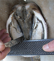
| Fig. 13. A rasp is placed across the foot and a ruler is placed within the collateral groove of the frog to measure the distance between the deepest part of the groove and the plane of the solar surface of the foot. This simple technique can be used to approximate the sole depth (courtesy of Dr John Schumacher). |
there will generally be a substantial increase in the proportion of the toe relative to the heels. A long toe will also be accompanied by an increased distance between the toe and the apex of the frog. The sole-wall junction should be solid and compact. Widening or fissures in the sole-wall junction and hoof wall separations dorsal to the sole wall junction occur with lengthening of the toe. The healthy sole tends to be concave and callused adjacent to the sole wall junction (white line). It should have a gradual slope from the apex of the frog to the sole wall junction and not a significant "trough." The sole should be between 10 to 15 mm thick beneath the margin of the distal phalanx and should not deform when hoof testers are applied. A ruler calibrated in millimeters can be placed within the collateral groove of the frog to measure the distance between the deepest part of the groove and the plane of the solar surface of the foot. The consistent distance (10 to 11 mm) between the distal phalanx and the collateral groove depth at the apex of the frog allows the clinician to predict sole depth. If one imagines moving the ground plane proximally so that the distance from the ground plane to the depth of the collateral groove decreases, it becomes clear that sole depth decreases as collateral groove depth decreases
13 (Fig. 13).
Horses with poor heel structure typically have a poorly developed digital cushion and thin collateral cartilages. These soft tissue structures determine the overall conformation of the palmar portion of the foot. Clinicians should gain an appreciation for variation in the consistency and overall size of the digital cushion and collateral cartilages. The digital cushion can be palpated between a thumb placed between collateral cartilages and the fingertips placed on base of the frog. A sense of "normal" can be acquired by palpating the digital cushions of sound horses with "good feet" and comparing those findings with those of horses with poorly conformed feet. The depth of the combined tissues of normal digital cushion and frog should be approximately 2 inches, but this can vary among different breeds.13 Horses with under-developed digital cushions typically have low or under-run heels that lack stability and can be easily distracted independently or they may have contracted heels and narrow, non-weightbearing frogs.19,20
9. Conclusions The clinical examination of the equine foot has been well-described and is generally performed in lameness cases. Evaluation of the hoof capsule during the lameness examination will provide additional information as to the etiology and treatment of the lameness but will also serve as a guideline to apply therapeutic farriery and other preventive measures to maintain a healthy hoof. The morphology of the hoof capsule reveals deformation and changes in growth that occurs after increased or reduced force. The relationship between the limb and the foot indicate conformations that predisposes the foot to abnormal weight-bearing. Inversely, with the use of abnormal distribution of forces and the subsequent hoof capsule distortion as a template, appropriate farriery or therapeutic farriery will form at least part of the treatment plan. Here it is essential to be familiar with the biomechanics of the foot and how these forces can be altered to change the distribution of forces or the focal stresses on a given section of the foot.
References - Parks AH. Form and function of the equine digit. Vet Clin N Am Equine 2003;19:285-307.
- Parks AH. Aspects of functional anatomy of the distal limb, in Proceedings. Am Assoc Equine Pract 2012;58:132-137.
- Johnston C, Back W. Hoof ground interaction: when biomechanical stimuli challenge the tissues of the distal limb. Equine Vet J 2006;38:634-641.
- Eliashar E. An evidence based assessment of the biomechanical effects of the common shoeing and farriery techniques. Vet Clin N Am Equine 2007;23:425-442.
- Parks AH. Therapeutic trimming and shoeing. In: Baxter GM, editor. Adams & Stashak's Lameness in Horses. 6th edition. West Sussex: Wiley-Blackwell; 2011:986-992.
- Turner TA. The use of hoof measurements for the objective assessment of hoof balance, in Proceedings. Am Assoc Equine Pract 1992;38:389-395.
- Redden RF. Hoof capsule distortion: understanding the mechanisms as a basis for rational management. Vet Clin N Am Equine 2003;19:443-462.
- O'Grady SE, Poupard DA. Physiologic horseshoeing: an overview. Equine Vet Edu 2001;13:330-334.
- Castelijns HH. The basics of farriery as a prelude to therapeutic farriery. Vet Clin N Am Equine 2012;28:316-320.
- O'Grady SE, Castelijns, HH. Sheared heels and the correlation to spontaneous quarter cracks. Equine Vet Edu 2011; 23:262-269.
- O'Grady SE. How to manage sheared heels, in Proceedings. Am Assoc Equine Pract 2005;51:451-456.
- Turner TA. Examination of the equine foot. Vet Clin N Am Equine 2003;19:309-332.
- Schumacher J, Taylor D, Schramme MC, et al. Localization of pain in the equine foot emphasizing the physical examination and analgesic techniques, in Proceedings. Am Assoc Equine Pract 2012;58:138-155.
- Turner TA. Predictive value of diagnostic tests for navicular pain, in Proceedings. Am Assoc Equine Pract 1996;42:201-204.
- Dabareiner RM, Moyer WA, Carter GK. Trauma to the sole and wall In: Ross MW, editor. Diagnosis and Management of Lameness in the Horse. 2nd edition. St Louis: Elsevier Saunders; 2011:309-319.
- Eliashar E, McGuigan MP, Wilson AM. Relationship of foot conformation and force applied to the navicular bone of sound horses at the trot. Equine Vet J 2004;36:431-435.
- Booth L, White D. Pathological conditions of the external hoof capsule In: Floyd AE, Mansmann RA, editors. Equine Podiatry. St Louis: Saunders Elsevier; 2007:224-252.
- Dabareiner RM, Carter GK. Diagnosis, treatment and farriery for horses with chronic heel pain. Vet Clin N Am Equine 2003;19:417-441.
- Bowker RM. The concept of the good foot: its evolution and significance in a clinical setting. In: Ramey P, editor. Care and Rehabilitation of the Equine Foot. Lakemont, Georgia: Hoof Rehabilitation Publishing LLC; 2011:2-34.
- Moyer WA, Carter GK. Examination of the equine foot. In: Floyd AE, Mansmann RA, editors. Equine Podiatry. St Louis: Elsevier Saunders; 2007:112-127.