Aspects of Functional Anatomy of the Distal LimbReprinted with permission from the American Association of Equine Practitioners. |
| Glossary of Biomechanical Terms Ground Reaction Force The ground reaction force is the force exerted by the ground on a body that is in contact with the ground. It is depicted as a vector that represents the sum of all individual forces on the surface of the body in consideration.
Distribution of Force Anywhere there is contact between the body and the ground, there is a force between the body and the ground. However, the force is not necessarily evenly distributed. For example, if a horse is standing on sand, the pressure is primarily distributed across the middle of the foot, including the sole and part of the frog. However, if the horse is standing on a flat, unyielding surface,most of the pressure is distributed around the perimeter of the foot at the interface of the ground and wall. Center of Pressure The center of pressure is that point through which the ground reaction force acts. Therefore,the center of pressure is the point about which the forces from all the different areas of contact are evenly distributed. It is also called the point of zero moment because it is that point at which all the moments created by forces on the object,in this case the horse’s foot, cancel each other out. The center of pressure is static only if a horse is standing still. When a horse is moving,the location of the center of pressure is dynamic.When the center or pressure is moved to one side of the foot, the bone and joints will be subject to increased compressive stress, the collateral ligaments to reduced tensile stress,and the lamellae to greater tensile/shear stress and vice versa. When the center of pressure is shifted in a palmar direction, tension in the deep digital flexor tendon is reduced, the weight bearing by the palmar hoof wall is increased, and vice versa. Moment (Torque) A moment is the tendency of force to cause rotation about an axis. It is calculated as the product of the length of the lever arm and the component of the force that is at right angles to the lever arm. If there are two equal but opposite moments acting around an axis, no movement occurs. |
2. Anatomy of the Hoof and Distal Interphalangeal Joint
The hoof is the integument of the foot, and as such,it is composed of three layers: the epidermis, dermis,and subcutaneous tissue. It is also divided into 5/6 regions: the limbic (perioplic), coronary, parietal (lamellar),solar, and cuneate/bulbar regions. The hoof capsule is formed by the stratum corneum of the epidermis of all these layers. The wall is formed by the stratum corneum of three layers-the limbic, coronary, and parietal-and these layers are called the stratum externum, stratum medium, and stratum internum, respectively. Each region of the hoof is highly specialized. The stratum medium of the wall is formed from tubular and intertubular horn. The structure, size, and density of the horn tubules vary with which zone of the wall they are located. The moisture content of the wall similarly varies, being drier more superficially and more hydrated in the deeper layers. The interdigitations of the lamellae are highly specialized and provide a very large surface area of contact between the epidermis and adjacent dermis. The frog is much softer than the wall and sole, and the underlying subcutaneous tissue is greatly modified to form the digital cushion. Therefore, the wall is well adapted to weight-bearing, the sole adapted to protecting the underlying soft tissues and weight distribution, and the frog and digital cushion adapted to permit expansion of the foot and participate in damping of vibrations.
The distal interphalangeal joint (DIPJ) is a complex joint with three articulations: (1) between the middle and distal phalanx, (2) between middle phalanx and the distal sesamoid (navicular bone), and(3) between the distal phalanx and the distal sesamoid.There is very little movement between the distal phalanx and the distal sesamoid,5 so they are frequently treated as one unit and will be so for the remainder of this discussion. The distal interphalangeal joint is a ginglymus joint. However, because the saggital groove on the middle phalanx is very shallow and the opposing ridge on the distal phalanx very low, it also permits significant rotation and movement in the frontal plane.
3. Aspects of Distal Forelimb Function in the Stationary Horse
In a standing horse, the weight (mass times acceleration of gravity) borne by the limb is supported by the ground, which opposes the weight with an equal and opposite force. This force exerted on the hoof by the ground is the ground reaction force (GRF).At rest, both of these forces are approximately vertical.The weight of the horse is not uniformly distributed across the ground surface of the foot. By using a complex pressure transducer system, the distribution of the GRF on the ground surface of the foot has been examined under various conditions.6It has been shown that in a shod horse, the weight is borne relatively evenly over the area that the shoe contacts firm ground. In a barefooted horse that has just been trimmed and is standing on firm ground, weight-bearing is increased compared to the untrimmed state and ground contact is present over the frog but is not necessarily evenly distributed around the perimeter of the foot. In a barefooted horse that has been at pasture and then stood on firm ground, the weight-bearing is primarily at the heel and toe. The pattern of weight-bearing at the toe has been shown to vary; it is either spread broadly across the toe from the toe-quarter junction on one side to the other or restricted to the toequarter junctions without any weight-bearing at the center of the toe. When a horse is placed on a surface that deforms to the shape of the foot, the weight-bearing area becomes much larger and is broadly distributed across the center of the ground surface of the foot.
The mechanical interaction between the horse and the ground is measured with force plates that do not differentiate between weight-bearing by different parts of the foot but renders a single value. It is represented as a vector (GRFV). Vectors have a direction and magnitude. This vector represents the summation of all the forces acting on the foot.Measurements made this way can be broken into three components representing the three orthogonal planes: vertical, craniocaudal, and mediolateral.As such, they have a point of action. This point of action is given several names: point of force, point of zero moment, and center of pressure. This article will use the latter because its meaning is more intuitive to most people. At rest, the vertical component of the GRFV is much greater than either of the two horizontal components.
The weight of the body borne by the limb is transmitted through the limb by the skeletal system.The question that arises is, how is this force transmitted from the skeletal system to the ground?Based on clinical evidence, it has been assumed that the lamellae suspend the distal phalanx within the hoof capsule. In horses with laminitis in which the lamellae are severely damaged, the distal phalanx displaces within the hoof capsule. Additionally, it is possible to remove the majority of the sole in a horse for therapeutic reasons, and the horse is able to bear weight on the wall without the distal phalanx displacing. How does this correlate with the fact that in a shod horse on a firm surface the weight is distributed around the periphery of the foot, but in a barefoot horse standing on a yielding surface the weight is not distributed around the wall but across the center of the foot?6 While it is not possible to measure where the force is going within the tissues of the hoof capsule and lamellae, this has been modeled with finite element analysis, which supports the intuitive position.7 When the weight is spread over the center of the ground surface of the hoof, it indicates that the forces associated with weight bearingare directed to the wall through the sole, and then through the lamellae.7
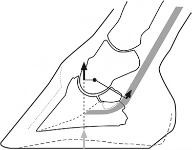 |
| Fig. 1. At rest, the ground reaction force (gray arrow) is dorsal to the center of rotation of the distal interphalangeal joint. As such, it creates an extensor moment that is opposed by an equal and opposite moment, the flexor moment, generated by the force in the deep digital flexor tendon so that the foot is stationary. |
The center of pressure varies between horses but is approximately in the center of the ground surface of the foot (Fig. 1). This is dorsal to the center of rotation of the distal interphalangeal joint. The force exerted through the skeletal system is acting through the center of rotation of the distal interphalangeal joint. Therefore, the GRF creates a moment about the distal interphalangeal joint. A moment is the tendency to cause rotation of a body about an axis. This moment created by the GRFV will cause the joint to dorsiflex (hyperextend) if unopposed;this moment is the extensor moment.In this case, the axis is the center of rotation of the distal interphalangeal joint. The magnitude of the moment is the product of the force and the length of the moment arm. The force is the magnitude of the GRFV. The length of the moment arm is the shortest distance between the line of action of the GRFV and the center of rotation of the DIPJ (i.e., the moment arm is perpendicular to the line of action of the GRF). Because the foot is in a stable position flat on the ground, the extensor moment must be opposed by an equal and opposite moment, which is the flexor moment. The flexor moment is the product of the force in the deep digital flexor tendon and the length of the moment arm, which is the shortest distance from the center of rotation of the DIPJ to the tendon.
4. Aspects of Distal Forelimb Function at the Trot
So far, this description has covered the dynamics of the foot of a horse standing at rest, but what about the foot of a horse that is walking or trotting? The stride is divided into flight and stance, and this discussion will be confined to the stance phase.At the beginning of the stance, the limb is fully protracted, with the foot out in front and the limbal most fully extended. After the foot comes to rest,the body continues to move forward and the trunk descends. As it does so, the metacarpophalangeal joint (MCPJ) dorsiflexes (hyperextends) and the distalinterphalangeal joint flexes; that is, they are rotating in opposite directions. At midstance, the limb is vertical and the MCPJ dorsiflexion and the DIPJ flexion have peaked. After midstance, the limb moves toward full retraction. As it does so,the MCPJ decreases (but is still dorsiflexed) and the DIPJ changes from flexion to dorsiflexion so that at the beginning of breakover, both joints are dorsiflexed.
As the limb moves through the stance phase of the stride, the GRFV changes and these changes reflect the different phases of the stride. The magnitude of the vertical component of the GRFV is very low immediately after the foot contacts the ground, increases as the horse bears more and more weight,and then decreases so that it is again very low when just before the foot leaves the ground. In the forelimbs,the horizontal component of the GRFV is, for approximately the first 60% of the stride, in the opposite direction to the movement of the horse, that is, it is a braking force.8 During the last 40% of the stride, the horizontal component of the GRFV is in the same direction as the movement of the horse, that is, it is a propulsive force.
The strains in the flexor tendons, accessory ligament of the deep digital flexor tendon, and suspensory ligament9 reflect the magnitude of the GRFV and the angulation of the metacarpophalangeal and distal interphalangeal joints. The strains in the superficial digital flexor tendon and the suspensory ligament are greatest at the point of maximal weight-bearing and maximal dorsiflexion of the metacarpophalangeal joint. The strain in the deep digital flexor tendon (measured proximal to its attachment to its accessory ligament) does not increase as much as that in the superficial flexor tendon or the suspensory ligament because as the metacarpophalangeal joint dorsiflexes, the distal interphalangealjoint flexes, that is, the tendency for the tendon to stretch around the metacarpophalangeal joint is offset, at least partially, by its tendency to shorten around the distal interphalangeal joint.The accessory ligament of the deep digital flexor is under greatest tension when both these distal joints are dorsiflexed but before the magnitude of the GRFV has decreased markedly.
Based on the kinematic and kinetic events of the stride, stance is divided into three phases.4 The first is the impact phase, which begins at first contact and is defined by the presence of shock waves present in the distal limb and is associated with landing and initial loading of the limb. The support phase begins at the end of the impact phase and ends at heel lift-off. It is the phase of the stride when the limb bears maximal load and the period before and after maximal loading. Its beginning is actually a continuation of the initial loading of the limb that begins at first contact but without the vibrations of impact. Its ending at heel-off signifies the kinematic event because the unloading of the limb continues until toe-off. Breakover begins with heel-off and ends with toe-off. The end of breakover is like the impact phase in that it is associated with shock wave vibrations, but they are of considerably lower magnitude.
The impact phase, which lasts 25 to 50 milliseconds,is further subdivided into two parts associated with two collisions.4 The first is the impact of the foot with the ground, which only lasts a few milliseconds followed by a second, which involves the impact of the weight of the body and limb of the horse with the foot. These two impacts overlap to some degree and set up a series of irregular shockwaves associated with the deceleration of impact.
The moments about the distal interphalangeal joint of a horse trotting are a function of the magnitude of the GRFV, the center of pressure, and the tension in the deep digital flexor tendon. Although the center of pressure at first contact is often at the heels or lateral quarter, because it moves very quickly toward the center of the foot and because the magnitude of the GRFV is low, this phase of the stride has received little attention. During the support phase of the stride, when the center of pressure is in a relatively constant position in the center of the foot and dorsal to the center of rotation of the distal interphalangeal joint, the extensor and flexor moment arms are relatively constant, and therefore the force in the deep flexor tendon directly reflects the magnitude of the GRFV. Towards the end of the support phase the tension in the distal portion of the deep digital flexor tendon and its accessory ligament increases so that the flexor moment increases. At the same time the magnitude of the ground reaction force is decreasing. Consequently,the center of pressure moves towards the toe, which lengthens the extensor moment arm such that the extensor moment equals the flexor moment and the foot remains flat on the ground. The heels lift off the ground when the flexor moment exceeds the extensor moment (which occurs because the center of pressure can move no further dorsally once it is at the dorsal margin of the toe).
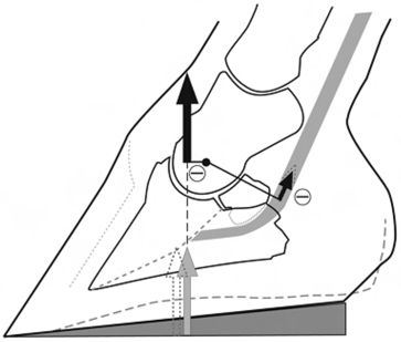 |
| Fig. 2. Elevating the heels (depicted at rest in this schematic)causes the simultaneous decrease in tension in the deep digital flexor tendon and movement of the center of pressure in a palmar direction toward the center of rotation of the distal interphalangeal joint. Thus, both moments are decreased equally, so there is no net movement of the foot (except for the initial elevation of the heels). The decrease in tension in the deep digital flexor tendon and the flexion of the distal interphalangeal joint cause the force on the navicular bone to be reduced. |
The distal limb has developed to absorb the energy associated with impact and the loading of the limb. It is known that impact vibrations are largely dampened by the time they have propagated to the proximal phalanx.10,11 The evidence indicates that the tissues that absorb the energy are the soft tissues of the hoof, for example, the lamellae and underlying dermis/subcutaneous tissue and the articular cartilage of the distal joints. Additionally,the structure of the digital cushion and the collateral cartilages and their associated venousplexuses suggest that a hemodynamic damping mechanism is present in the palmar half of the foot.12 The impact associated with loading of the limb by the weight of the trunk is also dampened by the combined action of the musculotendinous structures and the two distal joints in the limb, which, by extending the period over which the load is applied, decreases the maximum force on the distal limb. Dorsiflexion of the metacarpophalangal joint is associated with an increase in length of supporting tendons and ligament. The tendons are structured to store energy as they stretch and release it as they shorten, and the muscles are designed to dampen vibrations within the tendons.13 However, despite these protective mechanisms, excessive, repetitive strains can potentially damage tissues within the digit during either impact or loading.
5. Effect of Common Manipulations on Foot Function
The effect of shoeing horses on foot function is well documented. In short, it is known that nailing on steel shoes limits expansion of the foot 14 and causes the magnitude and frequency of impact vibrations to increase.10,15 It is also known that some shoe and pad combinations can ameliorate these changes.15
Two adjustments to shoeing commonly performed for horses with navicular disease are heel elevation and moving the point of breakover in a palmar direction.Our goal is usually described as taking the pressure off the navicular bone and making breakover“easier.” However, what do they really achieve in terms of the biomechanics discussed above? Elevating the heels causes the distal interphalangeal joint to flex. At rest, elevating the heels moves the center of pressure toward the center of rotation of the distal interphalangeal joint. Therefore, it shortens the moment arm of the GRFV, which means that the tension in the deep digital flexor tendon is reduced (Fig. 2). The reduction in the deep digital flexor tendon pressure tension in conjunction with the change in angle of the deep digital flexor tendon around the navicular bone substantially reduces the pressure on the navicular bone.16The biomechanics of the middle of the stride are similar with respect to the position of the center of pressure, but the magnitude of the GRFV and the tension in the deep digital flexor tendon are greater. Therefore, elevating the heels would be protective to the deep digital flexor tendon and navicular bone. However, moving the center of pressure in a palmar direction increases the load on the heels and increases intra-articular pressure. Additionally,flexion of the joint changes the distribution of pressure within the distal interphalangeal joint.Any of these effects of heel elevation are potentially deleterious.17
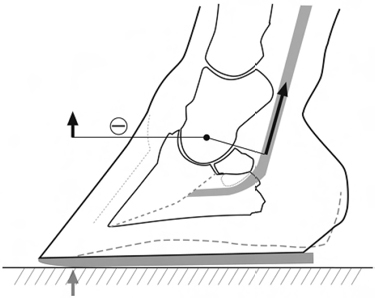 |
| Fig. 3. Moving the point of breakover in a palmar direction decreases the length of the moment arm at the beginning of breakover. This may cause breakover to occur slightly earlier, but it does not significantly reduce the duration of breakover or reduce the maximal force on the navicular bone. |
Moving the point of breakover in a palmar direction is thought to improve distal limb function in horses with diseases such as navicular syndrome.Moving the point of breakover in this manner does shorten the extensor moment arm at the initiation of breakover18 (Fig. 3). Because the flexor moment exceeds the extensor moment when the GRFV can no longer move dorsally, breakover may occur slightly earlier. However, it does not shorten the duration of breakover. Furthermore, it does not decrease the maximal pressure on the navicular bone as might be expected.18 This is because the pressure on the navicular bone is a function of the tension in the deep digital flexor tendon and the angle at which it bends around the bone. Therefore,maximal pressure on the navicular bone is a balance of decreasing tension in the deep digital flexor tendon as the load on the limb decreases toward the end of the stride and the increasing angle of the tendon around the navicular bone; the peak pressure on the navicular bone occurs at approximately 65% of the way through the stride,whereas breakover occurs at approximately 85% of stance.16,18 These findings would argue against the effectiveness of moving the point of breakover in a palmar direction for horses with navicular disease.However, it appears that rolling the toe smoothes out the breakover process.19
In horses with laminitis, in addition to raising the heels and moving the point of break over in a palmar direction, it is common practice to fill the space between the branches of the shoe with a synthetic polymer to promote weight-bearing by the sole.This procedure is also done for horses with other clinical conditions of the foot. Clinical evidence is highly varied, indicating that it appears to improve the lameness in some horses but worsens it in others.However, the scientific evidence behind what it does is minimal. This evidence, discussed above,suggests that it will distribute the force of weightbearing over a much wider area of the sole.6 However,finite element analysis suggests that movement of the quarters abaxially during foot expansion pulls at the margins of the sole, causing it to flatten out.7 Therefore, any device that limits movement of the sole in this manner may either limit foot expansion or place excessive strain on the white line. Intuitively, the thickness and quality of the sole horn may also affect how the foot tolerates this maneuver. However, much remains to be resolved about the effects of this procedure.
The farriery and veterinary professions have made significant advances over the last 25 years in treating many conditions of the horse's foot, but undoubtedly we still have a long way to go. This progress has received contributions from new scientific knowledge, but much of our progress has been the result of reasoning (sometimes good, sometimes bad) and experience. The author believes that the next step in our progress requires more deliberate thinking by clinicians about concepts such as moving the center of pressure and changing distribution of force into the development of therapeutic plans that involve biomechanical manipulation of the horse's foot.
References