Flexural deformities of the distal interphalangeal joint (clubfeet)Reprinted with permission from Equine Veterinary Education (EVE). Original published in Equine Veterinary Education Vol 27 April 2012.S. E. O'Grady Keywords: horse; flexural deformity; clubfoot; farriery; inferior check ligament desmotomy Summary A true clubfoot results from a flexural deformity of the distalinterphalangeal joint that is characterised by a shortening of the deep digital flexor tendon musculotendinous unit.Flexural deformities are a problem not only in foals but are also responsible for the clubfoot conformation seen in mature horses. Treatment is most successful when the cause is investigated and therapy initiated as early as possible, and when the biomechanical properties of the foot are thoroughly understood. Flexural deformities in foals and mature horses are addressed through appropriate farriery, often combined with surgery. Introduction Despite the recent advances in breeding, nutrition and farm management, flexural deformities are still a reasonably common occurrence. A flexure deformity can be defined as a shortening of the musculotendinous unit of the deep digital flexor tendon (DDFT) that results in hyperflexion of a given anatomic region of the limb (Adams 2000; Greet 2000; Hunt 2000, 2011; Greet and Curtis 2003). Flexural deformities have been traditionally referred to as 'contracted tendons'; however, the primary defect appears to be a shortening of the musculotendinous unit rather than a shortening of just the tendon portion; making 'flexural deformity' the preferred descriptive term (Kidd and Barr 2002).Shortening of the musculotendinous unit produces a structure of insufficient length to allow normal alignment of the distal phalanx (P3) relative to the middle phalanx and results in variable clinical signs ranging from an upright hoof angle to a clubfoot. The focus of this paper is to define and recommend therapy for flexural deformities involving the DDFT and the distal interphalangeal joint (DIPJ). The emphasis is on the forelimb unless otherwise stated in the text. The paper is divided into flexural deformities of foals and flexural deformities of mature horses. Severe flexural deformities of foals and mature horses are commonly referred to as clubfeet.
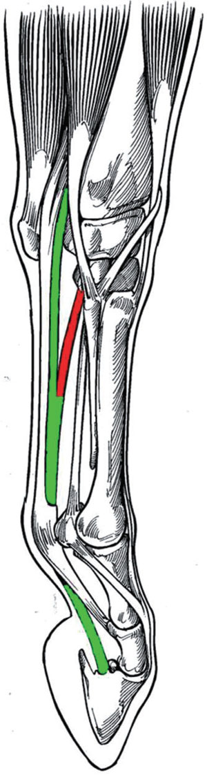
Anatomy review In the antebrachium, the muscle bellies of the DDFT lie directly on the caudal aspect of the radius and are covered by the muscle bellies of the superficial digital flexor tendon (SDFT) and the flexors of the carpus. The deep digital flexor muscle consists of 3 muscle bellies (the humeral head, the inconsistent radial head and the ulna head), which form a common tendon proximal to the carpus. This tendon, along with the SDFT, passes through the carpal canal and continues down the palmar aspect of the third metacarpal bone. Below the fetlock, at the level of the middle phalanx, the DDFT perforates the tendon of the SDFT, continues distally and inserts on the flexor surface of the distal phalanx (P3). A strong tendinous band known as the accessory ligament of the DDFT (AL-DDFT) originates from the deep palmar carpal ligament and fuses with the DDFT at the middle of the metacarpus (Fig 1). The design and function of the anatomical structures is such that any prolonged shortening of the musculotendinous unit affects the position of the DIPJ. The palmar surface of the distal phalanx is pulled palmarly by this shortened musculotendinous unit, placing the DIPJ in a flexed position. The alignment of the bone within the hoof capsule remains constant while the hoof capsule is pulled with the distal phalanx. The flexed position of the DIPJ combined with the altered load on the foot leads to a rapid distortion of the hoof capsule and thus the clubfoot conformation. It can also be noted from the anatomy that transecting the AL-DDFT, when necessary, lengthens the musculotendinous unit either functionally or by allowing relaxation of the proximal muscle belly associated with the DDFT. Classification of flexural deformities (clubfeet) Flexural deformities have been classified as type 1 where the hoof-ground angle is ≤90° and type 2 where the hoof-ground angle is >90° (Adams 2000). A recent method of classifying flexural deformities using a grading system (Grade 1-4) has been proposed (Redden 2003).Regardless of the method, it would appear beneficial to classify the severity of the flexural deformity to devise an appropriate treatment plan and monitor the response to a given therapy. A grading system would also enhance record keeping as well as improve communication between the veterinarian, farrier and owner with regard to treatment strategies. A Grade 1 clubfoot has a hoof angle 3-5° greater than the contralateral foot and a characteristic fullness present at the coronet. The hoof-pastern axis generally remains aligned. A Grade 2 clubfoot has a hoof angle 5-8° greater than the contralateral foot, the angle of the hoof-pastern axis is steep and slightly broken forward, growth rings are wider at the heel than at the toe, and the heel may not touch the ground when excess hoof wall is trimmed from the heel. A Grade 3 clubfoot has a broken-forward hoof-pastern axis,often a concavity in the dorsal aspect of the hoof wall, and the growth rings at the heels are twice as wide as those at the toe. A Grade 4 clubfoot has a hoof angle of ≥80°, a marked concavity in the dorsal aspect of the hoof wall, a severe broken-forward hoof-pastern axis, and the coronary band from the toe to the heel has lost all slope and is horizontal with the ground (Fig 2). For simplicity, the author uses a grading system based on the severity or degree of flexion noted in the DIPJ on the lateral radiographic projection to classify flexural deformities. Flexural deformities in young horses Flexural deformities in foals can be divided into congenital or acquired deformities. As such, congenital deformities are noted at birth, and acquired deformities generally occur during the first 6 months of life as the foal grows and develops. Congenital flexure deformities Congenital flexural deformities are present at birth,may involve a combination of joints (e.g. carpus,metacarpophalangeal and DIP joints) and are characterised by abnormal flexion of these joints and their inability to extend. Proposed aetiologies of congenital flexural deformities include malpositioning of the fetus in utero, nutritional mismanagement of the mare during gestation, teratogens in various forages ingested by the mare and maternal exposure to influenza virus, or the deformities could be genetic in origin (Kidd and Barr 2002;Hunt 2011). The affected foal tends to walk on the toe of the hoof capsule, is unable to place the heel on the ground and assumes a so-called 'ballerina' stance.Treatment of foals with a congenital flexural deformity varies with the severity of the deformity. A mild to moderate flexural deformity in which the foal can readily stand, nurse and ambulate is generally self-limiting and resolves without treatment. Brief intervals of exercise once or twice daily in a small paddock on firm footing for the first few days of life may be all that is necessary for the deformity to resolve. If the condition is severe or has not improved by the third day post foaling, i.v. administration of oxytetracycline (2-3 g) repeated every other day if necessary is frequently beneficial (Madison et al. 1994). A variety of bandaging techniques and splints are used, along with physical therapy, to 'stretch' the involved area to hasten recovery. Foals with severe congenital flexural deformities usually do not have just one isolated structure or joint that is responsible for the deformity, therefore, in the author's opinion, the use of a toe extension is not indicated. Acquired flexural deformities Acquired flexural deformities generally develop when the foal is aged 2-6 months and generally involves the DIPJ initially. The aetiology of this deformity is unknown, but speculated causes include genetic predisposition, improper nutrition (i.e. overfeeding, excessive carbohydrate [energy] intake, unbalanced minerals in the diet) and excessive exercise. A recent study looked at grazing patterns in a small number of foals and showed that foals with long legs and short necks had a tendency to graze with the same limb protracted (van Heel et al 2006). Fifty percent of the foals developed uneven feet with a higher heel on the protracted limb, leading researchers to feel there may be a possible correlation between conformational traits and an acquired flexural deformity. It is the current author's opinion that a large contributing factor to this syndrome is contraction of the muscular portion of the musculotendinous unit caused by a response to pain, the source of which could be physeal dysplasia or trauma from foals exercising on hard ground.Discomfort may follow aggressive hoof trimming where excessive sole is removed, rendering the immature structures within the hoof capsule void of protection and susceptible to trauma and bruising. Any discomfort or pain in the foot or lower portion of the limb coupled with reduced weightbearing on the affected limb appears to initiate the flexor withdrawal reflex, which seems to cause the flexor muscles proximal to the tendon to contract, leading to an altered position of the DIPJ. This shortening of the musculotendinous unit shifts weight bearing to the dorsal half of the foot causing a decrease in sole depth and bruising of the sole, reduced growth of the dorsal aspect of the hoof wall, and excessive hoof wall growth at the heel to compensate for the shortening of the musculotendinous unit. As the flexural deformity may be secondary to pain in these cases, it is essential that the source of pain should be carefully evaluated by physical examination and by localisation using regional analgesia and diagnostic imaging techniques. A genetic component must also be considered for acquired flexure deformities, as some mares consistently produce foals that develop a flexural deformity in the same limb as the dam or grand dam in which a similar deformity is present. The genetic component of the flexural deformity may be the ultimate determinant of the severity of the deformity. A genetic component should also be considered in the aetiology of acquired flexural deformities, although there is no scientific evidence for this at present. Some mares appear to consistently produce foals that develop flexural deformities, sometimes in the same leg as was affected in their dam or grand dam. Mild acquired flexural deformities Clinical signs The initial clinical sign of a flexural deformity may be abnormal wear of the hoof at the toe, which is often discovered by the farrier during routine hoof care. Closer investigation may reveal that the dorsal hoof wall angle is increased and that after the heels of the hoof capsule have been trimmed to a normal length, the heels may no longer contact the ground. A prominent coronary band may or may not be present at this stage. Most foals affected to this degree have a mildly broken-forward hoof-pastern axis. Increased palpable digital pulse, heat in the affected foot, and signs of pain when a hoof tester is applied to the dorsal aspect of the toe are not uncommon clinical findings. These findings are generally the result of trauma or excessive weightbearing on the toe.
Treatment Conservative treatment, such as correcting the nutritional status of the foal (i.e. weaning the foal to avoid possible excessive nutrition from the mare), restricting exercise to reduce trauma, judiciously administering a nonsteroidal anti-inflammatory drug (NSAID) to relieve pain, administering oxytetracycline to facilitate muscle relaxation, and carefully trimming the hoof is, in the author's opinion, a good starting point. NSAIDs should be administered short-term and should be used judiciously in foals due to the potential side effects, such as gastroduodenal irritation and nephrotoxicity. For analgesia,the author will administer firocoxib (0.1 mg/kg bwt q. 24 h) or flunixin meglumine (1.1 mg/kg bwt q. 24 h) combined with a gastric protectant. Hoof trimming begins with lowering the heels from the middle of the foot palmarly until the hoof wall at the heels and the frog are on the same plane. The bars should be thinned or removed, and the heels adjacent to the sulci should be angled to 45° to promote spreading.Breakover is moved palmarly by creating a mild bevel,with a rasp that begins just dorsal to the apex of the frog and extends to the perimeter of the dorsal aspect of the hoof wall (Fig 3). If improvement is noted, this trimming regime is best performed at 2 week intervals. If the toe is constantly being bruised or undergoing abscessation, a hoof composite (Equilox or Vettec) can be applied to the dorsal aspect of the sole and the distal dorsal aspect of the hoof wall to form a toe 'cap' to provide protection.The acrylic composite-impregnated fibreglass or urethane composite used to form the toe cap covers the solar surface of the foot to the apex of the frog, protecting that area from further damage. A bevel toward the toe can be created is the composite with a rasp or Dremel tool to facilitate breakover. If there is adequate integrity of the dorsal hoof wall, the author believes that application of a toe extension is unwarranted. The above treatment can be temporary, appears to work best when initiated at the first sign of foot deformity before a marked flexural deformity is noted and, if possible, following elimination of inciting causes. Severe acquired flexural deformities (clubfeet)
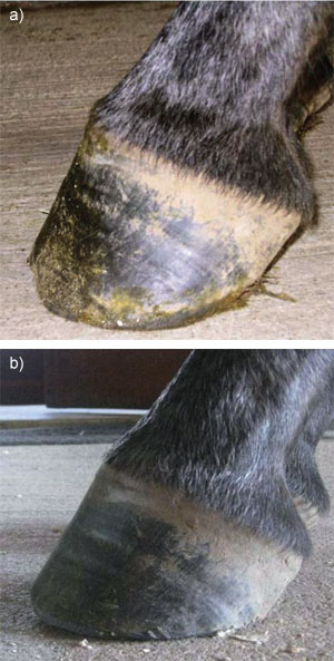
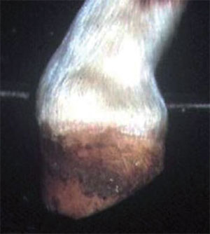
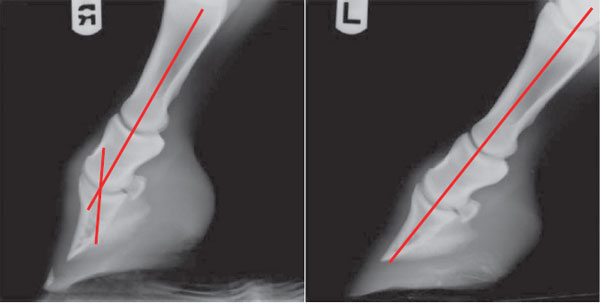
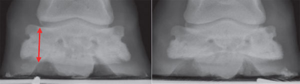
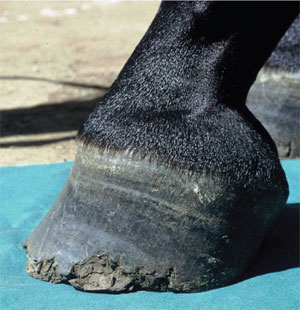
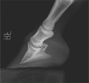
Clinical signs A mild acquired flexural deformity may progress in severity despite conservative treatment, or a severe acquired flexural deformity may be acute in onset.Asevere acquired flexural deformity is characterised by a foot with a hoof angle >60°, a prominent fullness at the coronary band, a broken-forward pastern axis, disparity in the length of the heel relative to the toe of the hoof, and heels that fail to contact the ground (Fig 4). If the cause of the deformity is allowed to persist, the foot eventually assumes a boxy,tubular shape due to the overgrowth of the heels to accommodate the lack of ground contact and approach the length of the toe. Increased stress on the toe will eventually cause a concavity along the dorsal surface of the hoof wall. Stress exerted on the sole-wall junction in the toe area will cause it to widen allowing separations to occur. The diagnosis is straightforward and based on the characteristic foot and limb conformation. Radiographs should be used to confirm the diagnosis and assess changes in the joint. The author will administer mild sedation (half the recommended dose of xylazine[0.33-0.44 mg/kg bwt i.v.] combined with butorphanol[0.022-0.066 mg/kg bwt] i.v.) and place each of the foal's feet on separate wooden blocks of equal height, which allows normal loading of both forefeet. Lateral-to-media land weightbearing (horizontal) dorso palmar views of both forefeet should be obtained. The degree of flexion of the DIPJ, the angle of the dorsal hoof wall and abnormalities at the margin of the distal phalanx should be observed(Fig 5). Interestingly, in all the cases where the author has obtained weightbearing (horizontal) dorsopalmar views, the width or thickness of the distal phalanx of the affected foot appears to be increased (Fig 6). This finding may possibly be due to radiographic projection with the change of angle of the distal phalanx to the horizontal but the author has also noted the change in width in foals without a flexural deformity. The significance of this finding is speculative but certainly adds credence to the genetic factor in the aetiology and the susceptibility of certain individuals to acquire a flexural deformity.
Treatment When a severe flexural deformity is present and confirmed during radiographic examination of the feet, conservative treatment and hoof trimming alone are generally of little benefit. Elevating the heels has been advocated,after trimming the foot, to reduce tension in the DDFT and to promote weightbearing on the entire solar surface of the hoof. Although elevating the heels improves the hoof-pastern axis and makes the foal more comfortable initially, the author has not been able to subsequently lower the heel or to remove the wedge and establish a normal hoof angle with the heel on the ground. Once a marked flexural deformity of the DIPJ and distortion of the hoof capsule are present or occurring, the author recommends transection of the AL-DDFT combined with the appropriate farriery.
Desmotomy of the AL-DDFT The author has been consistently successful in treating foals with severe flexural deformities with desmotomy of the AL-DDFT combined with the appropriate farriery. The author no longer uses a toe extension at the time of surgery, but applies an acrylic composite beneath the dorsal aspect of the toe to create a reverse wedge. The wedge affords protection for the toe region, appears to redistribute the load to the palmar aspect of the foot,increases the stresses on the DDFT, and restores the concavity to the sole. The author has found it beneficial to do both procedures at the same time while the foal is anaesthetised. The foal is placed under general anaesthesia and the surgery is performed in a routine manner as described elsewhere (Fackelman et al. 1983;Wagner et al. 1985; White 1995; Kidd and Barr 2002; Auer 2006). The farriery can be performed before or after the surgery. The heels are lowered from the point of the frog palmarly, until the sole adjoining the hoof wall (sole plane)at the heels becomes solid. Any concavity in the dorsal aspect of the hoof wall is removed with a rasp. The ground surface of the foot dorsal to the frog and the perimeter of the dorsal hoof wall are prepared for a composite using a rasp or Dremel tool. Deep hoof wall separations in the sole-wall junction at the toe are explored and filled with clay, if necessary, to prevent infection beneath the composite. Foals undergoing this procedure are usually aged 3-5 months, and so, because of their size and weight,reinforcing the composite with fibreglass is necessary. A small section of fibreglass is separated into strands and mixed with the composite. The composite is applied to the solar surface of the foot beginning at the apex of the frog and extending to the perimeter of the hoof wall where a thin lip is formed. The composite is moulded into a wedge starting at 0° at the apex of the frog and extending to 2-3°at the toe (Stone and Merritt 2009) (Fig 7). If desired, apiece of 3 mm aluminium plate can be cut out in the shape of the dorsal aspect of the sole. Multiple holes are drilled in the plate and it is gently placed into the composite. The aluminium is pushed down so that the composite material extrudes through the holes and the aluminium plate is then covered with additional composite. This additional reinforcement allows the older foals to be walked daily or turned out in a small paddock without the composite wearing out. Aftercare The surgical aftercare is at the discretion of the attending clinician. Controlled exercise in the form of daily walking or turn-out in a small paddock is essential. There is the potential for pain with the initiation of exercise, requiring close monitoring of the foal and exercise should be increased sequentially. The foal is trimmed at roughly 2 week intervals, based on the amount of hoof growth with the objective of establishing normal hoof capsule conformation. The composite wedge is removed one month after the surgery. At subsequent trimmings, the heels are lowered as necessary and hoof wall at the toe is trimmed from the outer dorsal aspect of the hoof wall until the desired conformation is attained. No sole dorsal to the frog is removed. When the desired conformation is reached, the foot is trimmed in a routine manner on a monthly basis. It is important to emphasise that when the hoof capsule returns to an acceptable conformation, only that portion of the sole that is shedding should be removed to avoid any discomfort in the dorsal solar area that can result in the horse re-developing, to some degree, the original deformity.
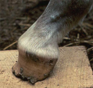
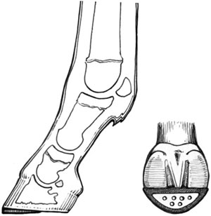
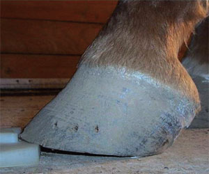
Flexural deformities in the mature horse Clubfeet There is minimal information in the veterinary literature regarding the management of a mature horse with a clubfoot. An upright conformation of the foot associated with a flexural deformity of the DIPJ is defined as a clubfoot (O'Grady 2011) (Fig 8). A flexural deformity is generally diagnosed and treated while the horse is immature but often a mild flexural deformity is ignored or the foal is treated inappropriately. When the horse enters training,the existing flexural deformity may become exacerbated by the type and amount of exercise, inadequate farrier care, such as inappropriate or infrequent trimming and shoeing, or some type of underlying disease. When a clubfoot conformation is acquired in the mature horse, it is almost invariably secondary to an underlying cause or disease, such as: an injury that results in a nonweightbearing lameness; excessive trimming of the toe resulting in solar pain; chronic, low-grade laminitis; or chronic heel pain. Furthermore, flexural deformities have been reported as a cause of decreased athletic performance and chronic, low-grade lameness in the mature horse (Balch et al. 1995; Turner and Stork 1998). The altered biomechanics of the foot result in an increased load (i.e. weightbearing) being placed on the dorsal section of the foot leading to decreased sole growth, sole bruising, a shortened stride on the affected limb, and various degrees of lameness and poor performance. The majority of horses with a clubfoot maintain soundness, yet the clubfoot conformation and the altered load on the foot may be responsible for poor performance. The hoof capsule distortion To apply the appropriate farriery, understanding the proposed mechanism leading to the clubfoot conformation is helpful. When a flexural deformity is present, the musculotendinous unit is shortened, the degree of which is dependent on the amount of flexion in the DIPJ. This causes a disparity of hoof wall growth, with more growth at the heel than at the toe to compensate for the decreased length of the soft tissue structures. The frog generally recedes due to the excessive hoof wall growth at the heels so that the energy of impact is assumed entirely by the hoof wall, bypassing the soft tissue structures in the palmar foot and transferring the load directly onto the bones of the digit through the laminar interface. The flexural deformity, combined with the excess hoof wall growth at the heels, places the DIPJ in flexion and distal phalanx in an abnormal alignment relative to the digit,promoting toe-first landing, and excessive load is assumed by the dorsal section of the joint and the dorsal section of the foot. Hoof abnormalities associated with clubfoot conformation are thin flat soles, poor hoof wall consistency(especially at the toe), toe cracks, hoof wall separation and 'white line disease' (O'Grady and Poupard 2003).Injuries associated with a high hoof angle are thought to include inflammation of the DIPJ due to abnormal loading of the joint, sole bruising and increased strain on the suspensory ligaments of the navicular bone (Turner 1992).
Radiology Good quality radiographs, consisting of lateral to medial and weightbearing (horizontal) dorsopalmar views, are necessary for the clinician and farrier to evaluate and treat a horse with a clubfoot. Good soft-tissue detail allows the distortion of the hoof capsule to be accessed. A lateral to medial radiographic examination reveals the weightbearing properties of the foot, the position of the distal phalanx within the hoof capsule, solar depth, length of the heels, the osseous integrity of the perimeter of the distal phalanx, and the severity of the flexural deformity of the DIPJ. The degree of flexion indicates the amount of shortening of the musculotendinous unit. The radiographs are used to diagnose any pathology present, determine treatment options and can be used as a template for farriery (Fig 9). Therapeutic farriery Therapeutic farriery forms the mainstay of treatment for clubfeet. Farriery should be based on principles rather thana particular method, and the principles remain the same regardless of the severity of the flexural deformity (O'Grady 2009). The principles are to achieve normal alignment between the proximal, middle and distal phalanges, and thus normal orientation and loading of the distal phalanx relative to the ground. Trimming and shoeing is aimed at removing weightbearing from the toe and dorsal aspect of the distal phalanx and re-establishing weightbearing to the entire solar surface of the distal phalanx and the corresponding hoof wall. Historically, farriers have been taught to trim (lower) the heels to correct the distorted hoof capsule and promote weightbearing in the heel area, but this type of trimming comes with a price. As the severity of the flexural deformity increases, so too does the shortening of the musculotendinous unit; therefore,lowering the heels directly increases the tension within the musculotendinous unit, and these stresses may lead to irresolvable tearing of the dorsal lamellae and widening of the sole-wall junction similar to that seen in the chronic laminitic hoof (Floyd 2007). The increased forces placed on the DDFT also promote hoof capsule distortion and abnormal loading.
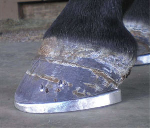
Farriery Distinguishing between a foot with steep hoof angle and a true clubfoot is important. High hoof angles without phalangeal misalignment or with mild phalangeal misalignment can generally be improved by gradually lowering the heels in a tapered fashion from the apex of the frog to the heels. This increases the ground surface of the foot and attempts to re-establish weightbearing on the entire solar surface of the foot. Breakover is moved palmarly at the same time to compensate for any increased tension in the DDFT created by lowering the heels. If improvement is noted, trimming should be repeated at 4 week intervals. Farriery to correct a high hoof angle accompanied by a flexural deformity becomes more of a challenge. Again, the object of farriery is to load the heels, compensate for the shortening of the DDFT and improve the hoof-pastern axis. To accomplish these objectives, farriery is directed at lowering the heels, but the amount to remove can be difficult to determine. In mild to moderate clubfeet, an estimate of how much heel to remove can be made by placing the thick end of a 2° or 3° wedge pad under the toe of the foot and allowing the horse to stand on it (Fig 10). If the horse does not resent the tension it places on the DDFT, this test allows the farrier to safely trim the hoof wall at the heels in a tapered fashion starting at the widest part of the foot using the thickness of the degree pad as a guide. The toe is shortened by trimming the outer surface of the dorsal hoof wall with a rasp. The trimmed foot is fitted with a shoe that has the breakover forged or ground into it starting just dorsal to the apex of the frog and tapering toward the toe to further decrease the stresses on the DDFT. With the more advanced cases of clubfeet, the heels should still be lowered to load the heels and unload the toe, but the addition of heel elevation following the trim is necessary to compensate for the shortening of the musculotendinous unit. The amount of heel elevation needed can be demonstrated following the trim by placing the trimmed foot on the ground 15-20 cm palmar to the contralateral limb. A space will generally appear between the heels of the foot and the ground. The author uses a wedge shoe or places a degree pad or a bar wedge between the heels of the foot and the shoe to compensate for the shortening of the muscle-tendon unit (Fig 11). This method allows the heels to be weightbearing but at the same time decreases the stresses on the musculotendinous unit. Creating breakover in the shoe to further relieve stress in the DDFT, as described above, is essential. Farriery combined with surgery In selected cases, horses with a severe flexural deformity or horses that have not responded to appropriate farriery and remain lame may benefit from a distal check ligament desmotomy (Turner 1992; Floyd 2007; O'Grady 2011). This release procedure, along with therapeutic farriery, allows realignment of the distal phalanx within the remainder of the digit and readily allows the accompanying distortion of the hoof capsule to be improved. The author believes that if this surgery is being contemplated, it should be performed early in the horse's athletic career, before there is a significant hoof capsule distortion and before radiographic changes involving the DIPJ and/or the margin of the distal phalanx become evident. In the mature horse, the surgery can be performed under general anaesthesia or with standing sedation/local or regional analgesia. In the standing horse, the heel should be elevated by taping a 12° wedge to the foot to decrease tension in the inferior check ligament/DDFT complex allowing the check ligament to be easily identified, separated and transected away from the cutaneous incision. The client should be forewarned that the surgery involves an extended recovery period, and a blemish or fibrous thickening at the surgery site isi nevitable due to the mature nature of the tissue. Caution is advised when performing the farriery that accompanies the surgery because the soft tissue structures within the hoof capsule and the digit have adapted/accommodated for the distortion of the hoof capsule.The author will trim the hoof in moderation and,according to information obtained from the radiographs, then apply two or three 2° wedge pads using either a shoe or a cuff. After surgery, the horse is walked daily, and a degree pad is removed every 7-10 days depending on the comfort of the horse. After 3 weeks, the horse is allowed turnout in a small paddock for an additional 3 weeks and then turnout in a larger area for 3-6 months before exercise is resumed. The cosmetic appearance of the limb is maximised by keeping the limb bandaged for the first 6 weeks. In a limited number of cases that the author has managed or consulted on, the benefits returning the horse to soundness have far outweighed the rehabilitation process being labour intensive (S.E.O'Grady, unpublished data). The author has not realised any benefit in applying a toe extension to the shoe in the mature horse following surgery. Many mature horses with a clubfoot frequently have damage to the dorsal lamina similar to that found in horses with laminitis, and, therefore,toe extensions may markedly exacerbate detrimental mechanical forces on the lamellae. Conclusion The clubfoot can be a significant cause of equine lameness and a challenge to the veterinarian and farrier. The clinician must recognise and understand the altered mechanics that are placed on the osseous structures within the hoof and on the hoof capsule that accompany a flexural deformity involving the DIPJ. This understanding allows the clinician to apply the appropriate treatment and appreciate subsequent improvement. Additionally, it is essential to look beyond the deformed foot to identify and remove, if possible, any underlying cause(s). As with most disorders,early recognition and intervention significantly increase the chances for a successful outcome. This is especially true when dealing with the young horse. Thorough physical examination, high quality radiographic imaging, familiarity with composite materials, and surgical competence are all essential to properly treat horses presented with a wide range of clubfoot abnormalities. Owners and trainers must be informed regarding the severity of any individual horse's flexural deformity, the treatment options, the expected outcome, and the aftercare. Knowledge, skill, and interaction between the veterinarian and farrier are necessary for a successful outcome when treating a horse with a flexural deformity, regardless of whether treatment is limited to farriery or combined with surgery. Acknowledgements The author would like to thank Drs Jim Schumacher and Liberty Getman for their invaluable help in critiquing this manuscript. References
|