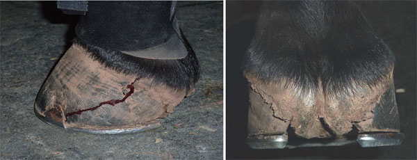
Sheared heels and the correlation to spontaneous quarter cracksReprinted with permission from Equine Veterinary Education (EVE). Original published in Equine Veterinary Education Vol 23 May 2011.S. E. O'Grady* and H. H. Castelijns† Summary The strong association between sheared heels and a spontaneous quarter crack is hard to ignore. Although inappropriate farriery may play a role, limb conformation and the landing pattern of the horse appear to be the dominant factors causing this type of hoof capsule deformation. The importance of determining the underlying cause and implementing the appropriate farriery cannot be over emphasised when managing a quarter crack associated with a sheared heel. The repair of spontaneous quarter cracks will be of little value, and the defect will have a tendency to recur, unless the cause is identified and rectified. Introduction Sheared heels as a clinical entity and a cause of lameness were first described in the veterinary literature 35 years ago(Moyer and Anderson 1975). A sheared heel is defined as a hoof capsule distortion resulting in a proximal displacement of one quarter/heel bulb relative to the contralateral side of the hoof (Turner 1992). The disparity between the lateral and medial quarter/heel bulb is generally 0.5 cm or more and is measured from the coronet to the ground or to the shoe. When the weight of the horse is not distributed uniformly over the entire hoof during the landing and/or weightbearing phase of the stride, one section of the foot, usually a heel bulb and accompanying quarter, receives a disproportionate amount of the total load. This repetitive disproportionate load causes the proximal displacement of the heel/quarter of the hoof capsule while the increased compressive stresses placed on the submural tissue in this area predispose the foot to various injurious conditions including a quarter crack (O'Grady 2002, 2005). While the diagnosis of a sheared heel is straightforward, the aetiology of the condition may be misleading and the farriery employed in the treatment is often based on opinions. Sheared heels appear to develop as an adaption-distortion of the hoof capsule as a consequence of limb conformation that results in an abnormal strike and loading pattern of the foot on the ground. Prevention or treatment of abnormal limb conformation is only possible in the foal; therefore in mature horses, therapy is directed toward managing the distortion of the hoof capsule. Spontaneous quarter cracks are a common cause of decreased athletic performance in competition horses and frequently lead to foot lameness (O'Grady 2001a; Moyer 2003; Castelijns 2006) (Fig 1). A true quarter crack originates at the coronet, extends distally through the full thickness of the hoof wall into the dermis, leading to instability, inflammation and/or infection. These cracks can be painful due to infection or, more commonly, the 'pinching' of the underlying dermis as a result of the movement of the unstable hoof wall. This 'pinching' occurs due to the vertical movement of the heel bulb and the outward movement of the entrapped ungual cartilage, axial to the origin of the quarter crack, during the loading of the foot. The recurrent nature of quarter cracks involving performance horses presents a challenging and often frustrating problem for equine veterinarians, farriers and horse owners, as these horses often need to continue to compete. Many causes of quarter cracks have been described,such as trauma to the coronet, pre-existing damage to the dermis from infection, abnormal hoof conformation, short shoes, inappropriate farrier practices or an abnormal landing pattern when the foot strikes the ground (O'Grady2001b). Yet the most consistent finding in all quarter crack cases is a foot conformation with a sheared heel on the side of the hoof with the defect and an abnormal strike pattern observed during the impact and loading phase of the stride. In fact, it is extremely rare to find a spontaneous quarter crack (as opposed to a crack due to outside trauma, such as a wire cut) that is not associated with this type of hoof capsule distortion. Various materials and techniques exist for stabilising and repairing hoof cracks, but none will be successful in the case of spontaneous quarter cracks, unless the cause of the hoof wall defect is determined and addressed through basic farriery, as these originate from the coronet, from the inside outwards(Moyer 1983; O'Grady 2001b; Moyer 2003; Castelijns 2006;McKinlay 2009). This paper will discuss the proposed aetiology leading to the hoof capsule distortion termed sheared heels and its correlation with quarter cracks, along with the farriery methods used in the authors' joint practices to address sheared heels.
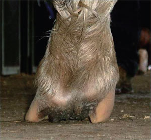
Structural changes to the foot The equine hoof capsule is a viscoelastic structure that has the unique ability to deform when weight is accepted uniformly (Parks 2003). However, if an unequal load is continually placed on one quarter/heel, over time, structural changes will become apparent. The increased load on one side of the foot causes the hoof wall to assume a steeper angle, i.e. the wall becomes straighter. This is a predictable response to increased load. Along with the increased hoof wall angle, other changes such as contracture of the heel subjected to the greater load will soon follow. The narrow heel will decrease the ground surface of the foot resulting in a lack of expansion on that side of the foot, making the solar surface in the palmar/plantar section of the foot asymmetrical. Over time, the hoof wall begins to 'roll under' on the affected side, which further decreases ground surface under that area of the foot. The side of the foot that first impacts the ground develops an outward flare due to bending of the hoof tubules (Fig 2). Over time, the stresses placed on the overloaded side of the foot exceed the ability of the hoof wall to deform and a distortion will occur (Parks 2003). This overload results in the coronet at the heel quarter and heel to be displaced proximally. Not only is the coronet displaced proximally but those structures located axially from the coronet to the middle phalanx are also displaced proximally and compressed, and this section of the foot becomes narrow(Fig 3). The submural tissue on the affected side may be subjected to excessive compressive forces that lead to stretching or tearing of the lamellae resulting in haemorrhage. It is thought that the exudation of fluid in the submural tissue increases pressure and will eventually disrupt the coronary corium contributing to the formation of a defect. Furthermore, a recent study of horses with quarter cracks, showed the free margin of the ungual cartilage above the coronet at the site of the crack to be <15 mm, as a result of the proximally displaced quarter/heel (Castelijns 2006). This lack of free margin appears to interfere with the abaxial expansion of the ungual cartilage when the foot is loaded, leading to increased pressure in the sheared heel and trauma to the adjacent coronet. Mechanism
The presence of a sheared heel indicates a disproportionate weight distribution over a section of the hoof that anatomically cannot resist the additional stresses without distortion or displacement. In this area, there is dorsal migration of the reflection of the wall at its junction with the bar and there are densely packed growth rings below the coronet. On gross dissection, the coronary groove, instead of being circular on a cross section,becomes disto-proximally elongated and narrow in the displaced quarter/heel. The narrower coronet produces a thinner hoof wall in this area (Fig 4). The growth rate around the circumference of the hoof is usually approximately uniform, but regional disturbances in growth rate can occur that will either increase or decrease growth. The position of the coronary band is related to the balance between hoof wall growth at the coronary band and the rate of migration of the hoof wall distally. Furthermore, the rate of migration of the hoof wall is a balance between an active process occurring in the lamellae to cause them to move distally and the force on the wall from the ground reaction force. Clinical evidence suggests that hoof wall growth is at least in part, if not predominantly, inversely determined by the force of weightbearing at the ground surface of the wall (A.H.Parks, personal communication 2010). If the rate of hoof wall growth exceeds the rate of migration distally, the coronary band displaces proximally. This appears to be the mechanism in horses with sheared heels/quarters. Tightly placed growth rings below the coronet coupled with slow hoof growth would suggest that the wall is forced proximally. Whether or not this is a real phenomenon suggested by clinical experience has not been confirmed in a scientific manner. Aetiology
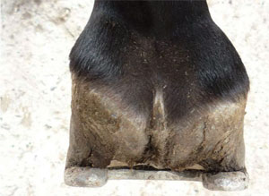
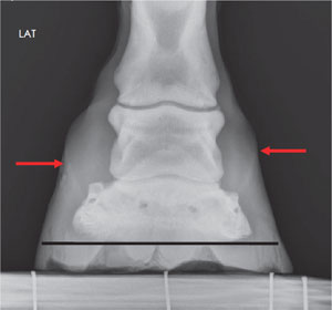
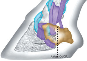
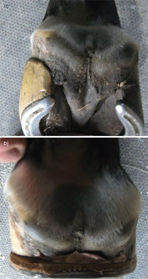
In order to formulate a rational approach to management,it is necessary to discuss the aetiology of sheared heels. The presence of a sheared heel when a spontaneous quarter crack occurs provides ample evidence that this type of hoof capsule distortion plays a role in the cause of the defect. It was assumed for years that inappropriate farrier practices may lead to this type of hoof capsule distortion when trimming methods, such as leaving the heels long or excessively lowering one side of the foot, would result in excessive forces/stresses being placed on a given section of the foot. The term used to describe this type of hoof capsule distortion was a lateral medial imbalance.Although this may indeed contribute, in the authors' experience, it does not predominately influence this type of foot conformation. To substantiate this theory, one author (S.E.O.) reviewed 50 dorsopalmar 0° radiographs on horses that had a foot with one heel bulb displaced proximally ≥0.5 cm. In all cases, it was clearly shown that the solar surface of the distal phalanx was approximately horizontal (parallel) with the ground. There was also an appropriate amount of sole depth under both the lateral and medial side of the distal phalanx. This would indicate that the disparity in heel height was not originating from the hoof wall and sole located distal to the distal phalanx (Fig 5). Anatomically, the distal phalanx occupies the dorsal two-thirds of the hoof capsule while the majority of the space in the palmar/plantar foot is occupied by soft tissue (Fig 6). The displacement of the heel thus seems to occur palmar/plantar to the body of the distal phalanx in the section of the hoof comprised of soft tissue. Conformational faults in the upper limb that change the horse's flight phase of the stride appear to be the major factor leading to this type of hoof capsule distortion. When such faults occur, the altered flight pattern causes the horse to impact the ground with one side of the foot prior to full weightbearing on the contralateral side of the foot. In the conformationally predisposed horse, the horse will generally have a narrow chest and the carpus will be rotated laterally. When viewed from the front, although the entire limb faces outward, or in some instances, inward, the axial alignment of the limb from above the carpus to the ground surface of the foot forms a straight line indicating a rotational deviation of the limb. For example, with a lateral rotational deformity, the knee faces outward, this moves breakover to an outward or lateral direction,thus altering the flight phase of the stride such that the foot is unable to land under the horse evenly on both heels. As the limb approaches the landing phase of the stride, this flight pattern forces the foot to contact the ground on one side and then sustain excessive load on the contralateral side. There is a far greater incidence of a sheared heel occurring on the medial side of the hoof but sheared heels on the lateral side are not uncommon. Using a slow motion video camera, one can actually distinguish the point where the foot impacts the ground on one side and the point where the hoof loads the surface on the other.Spontaneous quarter cracks generally occur directly above the point of maximum load and the largest force generated within the hoof wall. This point can be readily observed as there will be a focal proximal displacement in the coronet located directly above the quarter crack(Fig 7). Furthermore, there appears to be a correlation between an offset distal phalanx and sheared heels. Most commonly the distal phalanx is offset laterally within the hoof capsule rather than being directly under the proximal and middle phalanges causing the medial side of the hoof capsule to assume more load. Observation The evaluation of sheared heels begins with visual assessment of the hoof and limb conformation with the horse standing on a hard level surface. The gross changes noted in the foot are proportional to the amount of continual load sustained, the extent of structural damage and the duration of the condition. When sheared heels are present, the heel bulb on the affected side is displaced proximally and the structures above the heel bulb will be compressed when viewed from behind the horse. When viewed from the front, the hoof wall on the affected side is straighter and, in chronic cases, will begin to roll under the foot. There is generally a marked flare of the hoof wall present on the side opposite the affected heel in the toe quarter. When viewed from the affected side, the coronary band is displaced proximally above the damaged heel and will assume a horizontal contour, or a focal displacement, instead of having a gradual uniform slope from the toe to the heel. The solar surface of the foot reflects changes elsewhere in the hoof capsule: the foot will be less symmetrical; the sole in the quarter and heel area will appear wider on the side with the flare and narrower on the side with the sheared heel.
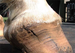
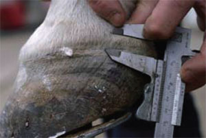
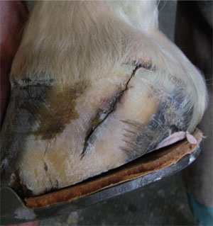
The vertically displaced heel is often affected by recurrent spontaneous quarter crack. When this occurs,the free margin of the ungual cartilage is usually diminished in feet with sheared heels due to upward displacement of the hoof wall at the heel. This is an important parameter to keep in mind when addressing a quarter crack. On palpation and measurement of a foot with a sheared heel and a spontaneous quarter crack, it is not uncommon to find the proximal border of the ungual cartilage at or below the coronary band. When a quarter crack is present, palpation of the ungual cartilage and moving the cartilage outwards (abaxially) by hooking a finger axially to it, tends to elicit pain and opening of the proximal margins of the crack (Fig 8). The painful reaction is usually not elicited on the contralateral side (to the sheared heel) by the same manipulation. The proximal margin of the quarter crack is always near the highest point of the vertical distortion of the coronary band when observed from the side. Measurement of the free ungual cartilage margin above the quarter crack by means of metric calipers reveals that when spontaneous cracks are present, this distance is ≤15 mm (2-15mm)(Castelijns 2006) (Fig 9). It is important to view the horse in motion, again on a hard level surface from the front and rear. This should be done at a walk and a trot. When viewed from behind, this should determine which section of the foot is contacting the ground initially and which portion of the foot is receiving the impact. The direction of breakover should be noted when viewed from the front. As the human eye is incapable of observing events with a duration of <0.02 s,and foot landing and loading can be quite different at different speeds and gaits, slow motion review of high speed film is highly recommended.
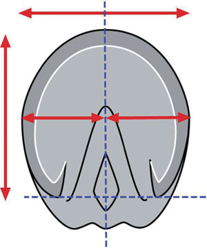
Farriery Farriery is directed toward unloading the hoof wall and decreasing the forces on the displaced side of the foot with the quarter crack. This is accomplished by improving the shape of the hoof and the landing pattern and the application of the appropriate shoe. When a horse develops a full thickness quarter crack, it is advisable to take the animal out of training to allow healing, but this is not always an option with competition horses. Constraints may be placed on the farriery due to the training and competition schedule of the horse. For example, the author (S.E.O.) likes to remove the shoes and stand the horse on a hard surface for 12-24 h prior to trimming and shoeing. This alone allows the affected side of the foot to settle into a more acceptable conformation (Fig 10). If a severe sheared heel hoof capsule distortion is present, the unshod foot can be stood on some form of frog support and the foot is placed in a soak bandage for 24 h (Snow and Birdsall 1990). This results in a profound change in hoof shape and the distance between the coronet and the middle phalanx will widen. If a quarter crack is present, when possible, the authors prefer to perform the farriery and then wait for the coronet to settle into a more acceptable position or slope before any type of repair is considered. If the repair has to be performed immediately, due to the competition schedule, the defect will be repaired with the coronet in a displaced position. Farriery is initiated by removing the shoes and again observing the horse walking on a hard surface noting the strike pattern of the foot. The authors use a double trimming method in an attempt to improve and unload the distorted quarter/heel. The foot is trimmed appropriately using the guidelines of a parallel hoof-pastern axis, the centre of rotation and heels of the hoof capsule trimmed to include the base of the frog (O'Grady 2009). To start, a line can be drawn across the widest part of the foot with a felt tip pen. The frog is trimmed to where it is pliable and the quarters and heels of the hoof capsule from the middle of the foot are rasped palmarly so that the heels of the hoof capsule and the trimmed frog are on the same plane if possible. An attempt is made to create as much ground surface under the affected heel as possible, which will often result in that side being marginally lower than the other side of the foot. The toe and quarters are reduced appropriately so when the trim is completed, the surface area on either side of the line drawn or the widest part of the foot will approximate each other (Fig 11). Lowering the heel on the displaced side of the foot is logical as it is the taller heel and it increases the ground surface of the foot on that side. Following the trim, the horse is again walked on a hard surface and some improvement in the landing pattern should be noted.
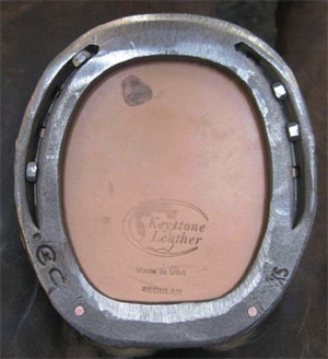
It is one author's opinion (S.E.O.) that, when initially managing a sheared heel, especially with a quarter crack,the horse should be placed in a bar shoe if possible. Bar shoes effectively increase the surface area of the foot, allow the palmar/plantar section of the foot to be unloaded, and decrease the independent vertical movement at the bulbs of the heels. The author's choice isa wide web steel straight bar shoe (Kerckhaert Shoes)1fitted symmetrically to the trimmed foot (Fig 12). Before applying the shoe, a second trim is performed under theproximally displaced quarter/heel, which goes from 0 mm at the ipsilateral toe (e.g. inside toe for medial sheared heel) to an average of 7 mm at the affected heel. The amount of heel that can be taken off in the second trim depends on the sole depth at the seat of corn and on the severity of the proximal displacement of the coronary band at the sheared heel. The amount of heel under the sheared heel that can be taken off with this second trim ideally corresponds to the difference in length/height between the 2 heels. Lowering the hoof wall at the quarter/heel will create a space between the shoe and the hoof wall on displaced side of the hoof (Fig 13). This improves the landing pattern, unloads the affected heel and allows the heel bulb to settle down and assume amore acceptable position. Feet with a low palmar/plantar angle rarely have enough sole depth under the affected heel for the second trim, in these cases the rest of the hoof wall can be raised with a rim pad or with a full leather pad and impression material. When a full pad is used,impression material (Equilox Pink)2 is placed in the palmar section of the foot from the apex of the frog palmarly except under the displaced heel where the second trim was performed. Only the first 2 nails should be placed in the toe of the shoe on the side with the sheared heel to effectively allow the displaced heel to settle into a more acceptable position. After the shoe is attached to the foot,the affected heel will rapidly descend onto the shoe,making the original space created by the second trim between the hoof wall and the shoe disappear. The extent of the second trim at the heel will determine the increase of free margin of the ungual cartilage above the coronet on the affected side of the hoof. This can be observed visually, palpated and measured with calipers. As most horses with a sheared heel have a predisposing limb conformation (e.g., a rotational deformity), these feet have a tendency to continue to deform the affected heel proximally and the double trim method usually has to be applied to some degree at each consecutive shoeing.Horses with this type of hoof conformation should be reset at 4-6 week intervals. Some cases will present with displaced heels with recurrent cracks and will resist lowering and widening of the sheared heel with the farriery methods described above. In these cases, one author (H.H.C.) treats the distortion of the hoof capsule at the site of the sheared heel with a full wall thickness sub coronary groove, applied with a rasp or Dremel3 tool about 20 mm below and parallel to the coronet. Care must be taken to go all the way through the wall to the laminar corium, from the end of the heel forward to the most dorsal part of the hoof distortion at the coronet. Horses may show discomfort from this procedure for a few days, especially if the laminar corium has been reached, which results in tiny pinpoint haemorrhage being visible. After the procedure, an antiseptic combined with a compressive bandage should be applied. The wall growth proximal to the groove will show a totally new, wider, abaxial direction as it is disconnected from the stresses being placed on the straight distal wall. Discussion The importance of determining the underlying cause and implementing the appropriate farriery cannot be over emphasised when managing sheared heels with quarter cracks. The strong association between sheared heels and a quarter crack coupled with limb conformation and the landing pattern of the horse is hard to ignore. The debridement, stabilisation and repair of spontaneous quarter cracks will be of little value, and the defect will have a tendency to recur, unless the cause is determined and rectified. Assessing the limb conformation, improving the foot shape and applying the appropriate trim/shoe appear to be as important as the repair technique used to stabilise the defect. Inadequate attention to these factors may account for the many failures encountered and the recurring nature of quarter cracks. The heels of the horse's foot have a relatively large amount of flexibility in the proximal to distal (vertical) axis.This can be explained by the anatomical features of the foot: a discontinuity of the hoof capsule between the heels with highly mobile structures interposed between which are the frog, digital cushion, venous and arterial plexa and fibro-cartilaginous connective tissue. In other words, although the dorsal wall is intimately attached to the parietal surface of the distal phalanx, the laminar attachment or suspension in the palmar/plantar section of the foot is far less rigid. This provides the flexibility necessary for function but also allows for proximal displacement of the heels when these receive excessive stress or a disproportionate load. Functionally this arrangement serves the bare-footed horse well as the hoof capsule at the heels is able to adapt to the uneven footing, but when shoes are applied this ability to adapt becomes modified. When trimming or shoeing modifications in the sagittal plane of the foot are being contemplated, it is important to be aware of this vertical mobility, and the tendency for vertical displacement of the heels. A wedge pad placed under the heels, for example, will cause proximal displacement of the heels (Castelijns 2006). The prognosis for sheared heels is good, provided a skilled, interested farrier is involved. It is also necessary to have a committed owner as these cases often require ongoing maintenance. Theoretically, the prevention and treatment of lameness and/or quarter cracks, caused by a hoof capsule distortion such as sheared heels is simple, but in practice it is often difficult to achieve. Being aware that there is a strong correlation between sheared heels and hoof wall problems, such as quarter cracks,makes prevention and treatment not only logical but imperative. Manufacturers' addresses
References
|