Examination of the Equine FootReprinted with permission from the American Association of Equine Practitioners. Originally printed in the 2010 AAEP Convention proceedingsAndrew Parks, MA, Vet MB, MRCVS, Diplomate ACVS 1. Introduction There are excellent journal articles and book chapters that describe the examination of the foot, some of which are listed as references.1-8 Reviewing them reveals that each clinician performs their examination in their own style, and they emphasize different aspects of the examination; however, they all have a method, and all describe an effective process to diagnose as effectively as possible the various conditions that affect the equine foot. This article describes the author's approach to examination of the foot, which is a synthesis of formal education received, personal experience, and experience of others. It is a reductionist rather than procedural approach. The majority of disease processes originating in the foot that cause lameness are associated with inflammation, usually related to trauma or infection. Disease processes associated with a marked focus of inflammation, typically associated with acute onset of lameness, are most likely to be identified with a basic examination, whereas disease processes associated with subtle symptoms and longer duration may require much more extensive examination of the limb and ancillary diagnostic tests. Furthermore, even the best examination in conjunction with ancillary diagnostic tests may not always obtain a definitive diagnosis, but the information gained may suggest an approach for symptomatic treatment. The latter is particularly important when access to advanced diagnostic technology, such as magnetic resonance imaging, is limited. Therefore, the following discussion is divided into two parts, the basic examination and a more detailed examination. The detailed examination is further divided into three main sections that provide different types of information. All examinations begin by gathering the presenting complaint, signalment, and history. With most foot problems, the presenting complaint is lameness. However, presenting complaint may also be the appearance of the foot. The signalment for any horse does not give specific information about the presenting complaint, but it does contain risk factors for certain conditions, which must then be correlated with information obtained from the history and physical examination. There are three main time points of importance in history taking: the date of the examination, the date that the problem was first noticed, and the date that the owner first knew/owned the horse. The second date gives an indication of the duration of the problem, and the length of time between the second and third dates gives the clinician an indication of how much history before this problem is known; this may lead to further enquiry about this time. No two sets of questions asked during a history taking are the same, because so many questions are predicated on the answer to a previous question; however, there is a common set of starting questions. After ascertaining the duration of the problem, questions should be directed at determining the nature of the onset-acute or chronic-and whether a specifically identifiable event can be linked to it. The clinician needs to know the progression of the disease, whether it has been constant, become worse, become better, or varied over the course of the history. Additionally, it is important to see if any treatment measures have already been taken and if so, what effect they have had. If the horse is able to work, what is the influence of exercise on the problem, and how does it change with the surface that the horse is worked on? Furthermore for feet problems, it is also important to determine the shoeing history if the horse is shod: when was the horse last shod, have there been changes in the shoeing technique, has the farrier changed before or during the course of the problem, etc. More general questions may need to be asked about the health and management of the horse, including questions about any other problems that the horse has had in the past, the husbandry of the horse, and disease-prevention management.
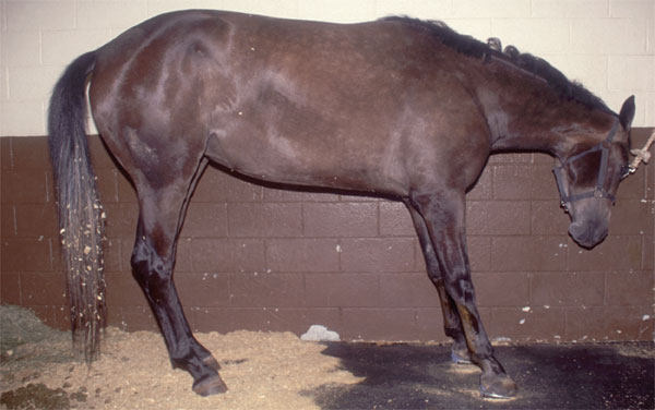
2. The Basic Examination 1. Examination of the Horse It is important to examine the horse as a whole and not rush to examine an extremity. The body condition of the horse should be assessed. Chronic pain related to lameness can cause weight loss. Pressure sores indicate that a horse has spent an excessive amount of time lying down. A horses posture may not only indicate which limb is lame, but it may also provide information about the nature of the problem, the classic example of which is the rockingback stance seen with bilateral forelimb laminitis (Fig. 1). If one heel is persistently held off the ground, it suggests that tension in a structure on the flexor surface is causing pain. 2. Basic Examination of the Foot Gross Examination of the Shape of the Foot It is important to have some appreciation for the size and shape of normal feet when conducting the examination, and although most observations on the size and shape of feet are subjective, values for the length and angle of the toe and the size of the foot in relation to the weight of the horse have been published. 1,9,10 The forefeet are more circular in shape when viewed from the ground surface, whereas the hindfeet may appear to be shaped like a diamond in which one end has been cut off.
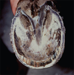
A visual inspection of the feet begins by walking around the horse and then looking at each foot more closely while it is on the ground. This should identify large defects in or distortions of the hoof capsule, scars, obvious swellings proximal to the coronary band, and mismatched feet. Then, the feet are picked up for examination of the ground surface of the foot, at which time changes in the loss of concavity and defects in the sole and changes in the width of and defects in the white line are noted (Fig. 2). In this manner, hoof-wall avulsions and heelbulb lacerations are readily identified. Likewise, marked concavity of the dorsal hoof wall in conjunction with a flattened or convex sole is almost pathognomonic for laminitis. Severe distortion of the hoof capsule proximodorsally is indicative of underlying proliferation of bone on the surface of the extensor process of the distal phalanx, often referred to as buttress foot. When in doubt about a questionable finding in one foot, examining the contralateral foot may determine whether or not it is abnormal. It should be borne in mind that some mild asymmetry between feet is within normal limits.
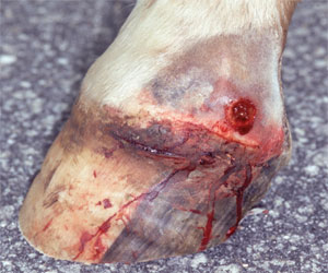
Swelling proximal to the coronary band may reflect a disease process in the pastern or within the foot. The distribution of swelling may be indicative of the nature of the problem. Circumferential swelling around the coronary band that extends up into the pastern is frequently associated with cellulitis. A small area of focal swelling at the coronary band is likely to be associated with an abscess that is about to spontaneously break open and drain. Inflammation of certain structures within the foot have characteristic patterns of swelling seen at or proximal to the coronary band. Inflammation of the distal interphalangeal joint results in dorsal symmetrical swelling, inflammation of the navicular bursa results in symmetrical swelling in the palmar aspect of the foot between the collateral cartilages, and inflammation of a collateral cartilage causes a unilateral welling proximal to the quarter of the hoof capsule (Fig. 3). The wall surface texture of the hoof capsule is normally smooth and therefore, appears shiny, but when it becomes roughened and irregular, it appears dull and may change to a lighter color. This usually reflects inflammation at the coronary band, although nutritional factors may potentially cause similar changes. It may involve all feet, one foot, or part of one foot. When it extends to the coronary band, it suggests that the problem is still active. If it is only one portion of the circumference of the foot, then the inflammatory process is localized. Because the wall migrates distally from the coronary band, the extent of the roughening proximodistally in relation to the length of the wall gives some indication of the duration of the process, including the date that it ceased if the inflammatory process is not currently active. Palpation Palpation of the foot is the most practical way to assess the temperature of the foot on a routine basis, and palpation of the palmar digital artery is performed to assess the pulse pressure (digital pulse), both of which are indicators of inflammation within the foot. The clinician should bear in mind that some variability in temperature of the hoof and strength of the digital pulse is normal; comparison with the other leg and repeated observations are helpful to confirm the importance of finding if in doubt. The pastern is palpated to detect possible surgical scars, and the palmar digital nerve is palpated for swelling and pain on deep palpation to detect a neuroma. The surface texture of the hoof and skin of the pastern is most appropriately assessed by feel, particularly the roughness of the surface of the hoof capsule and thickening or irregularity of the skin of the pastern. Palpation may also detect the presence of moisture on the foot and pastern, particularly at the hairline, that is not otherwise detectable. Examination of the coronary band may reveal a depression immediately proximal to the hoof capsule, which is indicative of distal displacement of the distal phalanx. Palpation is used to determine whether a swelling proximal to the coronary band is firm, edematous, or fluctuant. Digital pressure is applied, at first gently and then more firmly, to identify a focus of pain, if present. Structures proximal to the coronary band that may be palpated directly through the skin include the proximodorsal distal interphalangeal joint capsule, proximal aspect of the distal interphalalangeal joint collateral ligaments, proximal interphalangeal joint collateral ligaments, middle and proximal phalanges, common digital extensor tendon, deep digital flexor tendon and sheath, collateral cartilages, and digital cushion. Pressure may be applied to the navicular area through the deep digital flexor tendon and digital cushion. Deep sulci may need to be palpated for discomfort with the aid of a tongue depressor. The flexibility of the collateral cartilages may also be determined by palpation. Manipulation The distal limb should be flexed and extended to determine if it elicits a pain response or if there is a reduced range of motion. In general, structures that are associated with flexion/extension during normal locomotion, such as the tendons and their sheath, ligaments, joints, and navicular bursa, are likely to elicit a painful response. Usually, pain associated with the hoof capsule and non-articular portions of the distal phalanx are unlikely to do so. However, manipulation should be interpreted cautiously, because instability between two parts of the hoof capsule or instability between the hoof capsule and underlying distal phalanx may also be stressed by such handling. Rotation of the foot in relation to the remainder of the digit shows a remarkable degree of mobility in normal horses, which should not be interpreted as abnormal. Marked sprains of collateral ligaments might be expected to elicit a painful response after manipulation in this manner. Compression and Percussion Detection of pain within the foot is difficult with palpation because of the rigidity of the hoof capsule. Therefore, compression and percussion are used to identify and localize pain within the foot. Hoof testers are used to compress the foot. They should be applied in a systematic manner, typically starting at one heel, progressing around the quarter, toe, opposite quarter, and heel and followed by compression across both heels and from each side of the frog to the opposite heel. In addition to progressing around the foot, systematic application of hoof testers must also evaluate the sole at different distances from the white line. When the initial withdrawal response is mild, consistency with repetition and comparison with the contralateral limb is needed to determine if the response is clinically significant. In addition to eliciting a pain response, compression of the foot may also cause moisture to be expressed from defects in the hoof capsule and identify instability of fissures in the hoof capsule. Flexion of sole with hoof testers gives some indication as to its thickness. When applying hoof testers, it is frequently assumed that any pain response identified is coming from the solar aspect of the foot. Although this is frequently the case, it should be borne in mind that hoof testers might elicit a painful response from any of the tissues between their jaws. For example, when hoof testers are applied across the toe, the tissues affected include the integument of the wall as well as the integument of the sole and the distal phalanx. Percussion of the hoof capsule with a shoeing hammer is not performed as frequently as compression, but it occasionally yields information that hoof testers do not give. Additionally, it is useful when no hoof testers are available. Although the foot must be elevated off the ground to percuss the sole, the wall may be percussed with the foot on or off the ground, but the latter is easier. Paring With a Hoof Knife The ground surface of the foot provides clues regarding the quality of the sole, evidence of past trauma, and defects that are potential entry sites for infection. Defects in the ground surface of the stratum medium of the wall and the white line cannot be identified if a shoe is present. The manner in which paring is performed and when it is performed is related to the presenting clinical symptom. In horses that present with an acute marked onset of lameness in which an abscess is the most likely diagnosis, paring the foot is usually performed promptly. However, removal of shoes and paring of the ground surface of the foot is not recommended for horses with mild lameness and no obvious abnormalities of the ground surface of the foot until after the horse has been exercised and all pertinent diagnostic analgesia has been performed. Not all lameness originating in the foot requires paring of the sole, and when it is done, caution should be exercised to preserve the thickness of the sole whenever possible. In particular, if a horse is suspected of having laminitis, preservation of the sole thickness is very important, and therefore, paring should be very limited in extent or avoided. The frog may require trimming to expose the sulci or investigate primary diseases of the frog, such as thrush and canker.
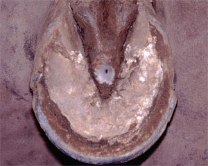
The ground surface of the foot in horses with marked lameness of abrupt onset is explored for puncture wounds and defects in the sole that are likely entry sites for infection. Both usually appear as dark marks. Naturally occurring defects are most likely to be present in the white line. Puncture wounds in the frog are frequently difficult to identify, because the elastic nature of the frog causes the entry wound to close over (Fig. 4); punctures in the collateral sulci are difficult to identify, because the exposure is poor. In horses in which the most likely diagnosis is an abscess, paring should be very focal and should extend through the full thickness of the hoof capsule until either purulent exudates or pinpoint hemorrhage is encountered. If the likely entry site is in the white line, it is preferable to explore the defect by enlarging the side adjacent to the wall rather than the sole, in effect creating a notch in the distal wall that extends proximally to the junction with the inner surface of the sole. In horses with milder but more chronic lameness, paring the sole is kept to a minimum. Lightly debriding of the surface of the sole frequently reveals hemorrhage related to bruising. Blood that extravasates into the sole maintains its red coloration, and it can further be distinguished from pigmentation, because it has a stippled pattern as it migrates down and around the horn tubules. Evaluation of Shoes and Shoeing The shoes should be evaluated to determine that the size of the shoe is appropriate for the size of the foot and that the type of shoe is appropriate for the type of work that the horse is performing. Uneven wear on the ground surface of the shoe may indicate areas of excessive weight-bearing. If a shoe has been on too long, it moves forward in relation to the heels and is likely to put pressure on the angles of the sole. Additionally, shoes that have been on too long may loosen and shift. 3. The Detailed Examination 1. Morphological Examination of the Hoof The goal of examination of the detailed morphology of the hoof is to identify deformation of the hoof capsule and changes in the growth pattern of the hoof that may indicate the presence of abnormal distribution of stresses within the foot. Increased stress or weight-bearing by a portion of the wall has three consequences that may be detected on physical examination: it may cause deviation of the wall outward or inward from its normal position, it may cause the wall to move proximally, or it may cause the growth of the wall to decelerate. A reduction in stress or weight-bearing, for the most part, has the opposite effect. In contrast to the situation where stress can retard hoof growth, there are occasions when hoof growth is accelerated in one part of the foot in relation to another; the best examples of this would be the growth pattern associated with inflammation or tumors. To accomplish a more detailed morphological examination of the foot, the foot should be viewed from all sides when it is on the ground, and then, the ground surface should be examined with the foot off the ground. Additionally, small changes in the shape of the hoof capsule may be better appreciated by careful palpation of the foot than by visual inspection.
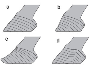
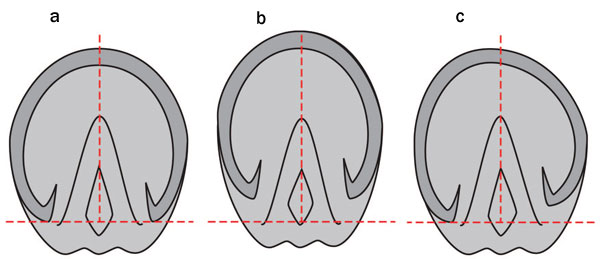
When the foot is viewed from the dorsal aspect, several abnormalities may be visible (Fig. 5). Flares or underrunning of the wall may develop at the quarters. The coronary band may be unevenly distributed. The most common development would be an even slope in the coronary band from one side of the foot to the other. However, more localized distortions of the coronary band may occur; a common example is that in which the coronary band in the median plane is distal to that at the toe-quarter junction. Examination of the growth rings may show divergence of the rings from one side to the other. The angle of the dorsal-horn tubules to the saggital plane should be noted; normally, they should be parallel, and therefore, when they appear tilted medially or laterally, it suggests that the whole hoof capsule may be tilted. When the foot is viewed from the lateral aspect, flaring of the toe and underrunning of the heels is readily appreciated. The coronary band should normally slope evenly from the toe to the heels. Evaluation of growth rings indicates a disparity in the growth of the heel and the toe, typified by the increase in heel growth and decrease in toe growth commonly seen in horses with laminitis (Fig. 6). However, regional irregularity in spacing of growth rings is not uncommon; the most frequently observed is a decrease in spacing at the quarter associated with proximal displacement of the coronary band. The heels are evaluated from the palmar aspect for their overall width and height. The heels frequently become narrowed when the foot itself is narrow. Additionally, the central sulcus may extend proximal to the hairline so that a cleft becomes apparent in the skin of the pastern between the heels. The overall height of the heels is readily assessed from the lateral aspect, but viewing from the palmar aspect is useful to compare the relative heights of the two heels; the classic example is the sheared heel in which one heel is displaced proximally in relation to the other. If a three-dimensional object changes in one plane, it will change in at least one other plane, and this is certainly true for the horse's foot. Therefore, examination of the ground surface of the foot reveals much about the changes in the wall of the hoof capsule (Fig. 7). In general, the frog is usually constant in length; its axis is almost always aligned with the medial plane of the foot, but its width is variable. Normally, the curvature of ground surface of the wall should be smooth, and it is almost symmetrical about the axis of the frog. The width of the ground surface should be approximately equal to its length, the maximal width is approximately halfway between the toe and heels, and the palmar margin of ground surface of the wall at its reflection is level with the base of the frog. Therefore, the contour of the wall can be examined in relation to its curvature and the position of the frog. These changes may affect the whole foot and therefore, are frequently symmetrical in the saggital plane, or they may be regional and therefore, usually asymmetrical. A narrow foot suggests that the toe is long or that foot expansion has become decreased, usually secondary to pain. In some individuals and some breeds, the toe is deliberately maintained long, but at other times, a long toe is inadvertent. A long toe will also be accompanied by an increased distance between the toe and the apex of the frog. When the ground surface of the heels are dorsal to the base of the frog, the heels are underrun and/or increased in length. If the contour of the wall is displaced away or toward the median plane in the dorsal two-thirds of the foot, this usually corresponds with a flare or underrunning of the wall, respectively. If only one heel buttress is displaced dorsally in relation to the base of the frog, it usually corresponds with the proximal displacement of that heel plus or minus the quarter-termed sheared heel.
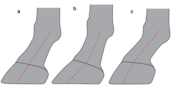
2. Examination of the Foot in Relation to the Rest of the Limb The structure of the distal limb is examined to identify features of that animal's conformation or balance that may contribute to undue stresses in any part of the foot, which may predispose to injury or disease. It is important that the structure of the distal limb is viewed both on and off the ground. When the distal limb is viewed standing on a level surface, except under unusual circumstances, weight-bearing forces the ground surface of the foot to be perpendicular to the pastern, and internal structures may have to accommodate for this. With the foot off the ground, the constraint of weight-bearing is not present, and the ligaments are relaxed. When the digit is viewed from the dorsal aspect with the foot on the ground, the pastern and foot should be in alignment (i.e., the median plane of the pastern and medial plane of the foot are parallel and in line with each other). If the median plane of the pastern appears to intersect the coronary band to one side of its center, it suggests that that side of the foot/hoof is elevated. When the digit is viewed from the side with the foot on the ground, the dorsal aspect of the pastern should be parallel with the dorsal hoof wall. This relationship is referred to as the hoof-pastern axis. When the hoof capsule forms a more acute angle with the ground than the pastern, the axis is said to be broken back, and when the angle is less acute, the axis is said to be broken forward (Fig. 8). This evaluation is somewhat subjective, because it depends on which part of the pastern is being compared with the hoof and the angle changes slightly with the phase of the shoeing cycle. The broken-back foot-pastern axis is associated with increased tension in the deep digital flexor tendon and greater force on the navicular bone during the stride, particularly at breakover. A brokenforward foot-pastern axis, usually associated with a flexural deformity of the distal interphalanageal joint, is thought to predispose to concussion of the dorsal sole.
Additionally, when viewed from the lateral aspect, position of the fetlock is related to the position of the ground surface of the foot. There is a traditional metric that states that an imaginary vertical line that bisects the metacarpus should intersect the ground at the point that the heels contact the ground. It is a function of the angle of the footpastern axis, the length of the pastern, and the size of the ground surface of the foot. Like other aspects of conformation, its significance is uncertain, but rationally, if the horizontal distance between the fetlock and foot is longer, it suggests that there is greater stress on the supporting ligaments and tendons. With the limb off the ground, the most common way to assess the relationship between the foot and the rest of the distal limb is to hold the metacarpus horizontal and sight along the palmar aspect of the limb. In this manner, the relationship between the ground surface of the foot and an imaginary line across the heel bulbs can be evaluated in relation to the axis of the limb. The ideal relationship frequently cited is that a line drawn across any two comparable points from the medial and lateral sides of the hoof capsule should be perpendicular to the axis of the metacarpus. This ideal relationship is thought to provide optimal distribution of weight within the foot and therefore, is frequently used as a guide for trimming the foot. Unfortunately, this relationship varies with rotation within the metacarpus and pastern and angulation at the metacarpophalangeal joint. Therefore, it is important that it is interpreted in conjunction with the appearance of the distal limb when placed on the ground and the morphology of the hoof capsule. 3. Examination of the Foot in Motion Observation of the distal limbs in motion should be performed at both a walk and trot, if possible, and should be performed from in front, to the side, and from behind the horse as it moves. It is only recently that advances in technology have allowed us to better understand the manner in which the foot moves at breakover, during flight, and on landing. It is now known that most horses land laterally at the heel or quarter and less commonly, land flat. Breakover is normally slightly lateral to or at the center of the toe. However, landing and breakover are rapid events that are difficult to discern by watching a horse at a trot, and therefore, they are best observed at a walk. A toe-first landing or medial first landing are both abnormal events at a walk and warrant further investigation. Additionally, excessive lateral-first or heel-first landing is likely to be abnormal. When mediolateral asymmetrical landing can be detected at a trot, it is likely to be significant. Although the implications of abnormal landing and breakover are not fully understood, it is most likely because of one or more of the following reasons. It could be because of the horse's conformation. It could be because of the horse's attempts to ameliorate pain in the foot. It could also be that the normal range of the stride is altered, usually because of a more proximal focus of pain, and therefore, an earlier or later breakover or landing may influence what part of the foot contacts and leaves the ground first. In addition to changing the way that the foot lands and leaves the ground, the horse may place its limb farther to or away from the median plane to redistribute weight, which would be accompanied by associated changes in flight after breakover or before landing. This is most likely to be seen when a horse positions its foot farther from the median plane to reduce lateral weight-bearing or vice versa. Flexion tests and toe/heel elevation tests are designed to stretch a set of structures before trotting to make an occult lameness apparent or a mild lameness more obvious. 4. Ancillary Diagnostic Aids There are three main objectives to ancillary diagnostic tests: exploration of an injury, usually with a flexible metal probe, diagnostic analgesia to localize the source of pain, and diagnostic imaging to identify discrete pathology. Their discussion is beyond the scope of this article. 5. Summary The manner in which the parts of the exam have been described is a reductionist approach because it is often easier to understand the significance of an individual finding in isolation. However, the examination is performed in the most clinically efficient manner so that several characteristics of the limb are being observed at one time. For example, when observing the distal limb on the ground from either the dorsal or lateral aspect, the morphology of the hoof capsule and the relationship between the foot and remainder of the distal limb when weight-bearing are evaluated concurrently. The basic examination is likely to provide a diagnosis for many common and simple disorders, such as a foot abscess. The detailed examination may, under some circumstances, provide a definitive diagnosis but is as likely or more likely to direct the clinicians attention to risk factors for injury and indicators of abnormal stresses. The three main areas that are assessed provide different types of information as described previously. In brief, the morphology of the hoof capsule reveals deformation and changes in growth that occur after increased or reduced force, and the relationship between the limb and the foot indicates conformations that may predispose to abnormal weight-bearing. Observing the limb in motion is most helpful to corroborate with findings identified when the horse is examined at rest; however, because there are limited data available for comparison of the landing and breakover patterns with different disease states, it is more of an art than a science at present. The correlation of the clinical findings to suggest a probable diagnosis may be good. However, at other times, the clinical findings may have conflicting implications; this may point to two separate clinical problems or a break in our understanding of the pathogenesis of the changes in structure of function observed. The latter is particularly important when attempting symptomatic treatment in the absence of a definitive diagnosis for whatever reason. In this instance, a measure of trial and error is warranted, and it is based on the preponderance of the evidence rather than the total evidence. Lastly, it should be remembered that, for some purposes, the hindfeet can be considered very similar in structure and function to the forefeet (for example, in the diagnosis of abscesses, avulsions, and distal phalanx fractures). However, when diseases such as those that can be attributed to hoof imbalance are considered, then the structure and function of the hindfoot should be considered separately from the forelimb. This is because the stresses associated with locomotion are sufficiently different (and less well-understood) that changes in the hoof capsule should not necessarily be interpreted in the same manner as they would be in the forelimbs. References
|