Guidelines for Trimming the Equine Foot: A ReviewReprinted with permission from the American Association of Equine Practitioners. |
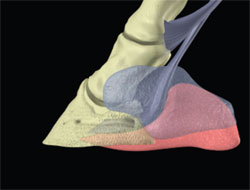 |
| Fig. 1. Relationship of the osseous structures (weight bearing) and soft-tissue structures (decrease concussion) of the equine foot. (Courtesy of Dr. Andrew Parks.) |
Function
Classically, the horse is thought to bear weight on the wall, bars, and immediately adjacent sole as well as the frog, to some extent. This seems true for a horse that is wearing shoes and standing on a flat,hard surface, but if the ground surface is soft and deformable, more of the ground surface of the sole and frog will assume a load-sharing function. Initial contact with the ground is generally made with the heels first, but many horses will land flat. Toe-first landing is considered to be abnormal and an indication of palmar foot pain.5 For clarity, the function or physiology of the foot will be considered during the impact and stance phase of the stride. The relationship of function to lameness is readily observed during these two phases of the stride. As the foot impacts with the ground, the physiologic process involves many structures simultaneously. The act of weight bearing is carried out by the hoof wall, adjacent sole, and partially through the frog and is then transferred through the lamina to the distal and second phalanx. The energy generated through impact is dissipated by the flexibility of the hoof capsule and the soft-tissue structures of the foot, especially the digital cushion.2,3,5 Flexibility of the hoof capsule, mainly caused by the expansion of the heels, is an interactive effort between the hoof capsule and the soft-tissue structures in the palmar foot. The exact mechanism by which the heels expand still remains to be documented. It is speculated that when weight is placed on the foot, the frog will flatten and spread abaxially to assist in absorbing the energy of impact. In fact, during impact, it seems that the second phalanx exerts pressure against the digital cushion, which forces this structure down on the frog and causes it to deform. The resulting force on the digital cushion causes it to expand outward, pressing against the ungual cartilages and spreading the quarters of the foot even more.2 Recent research has shown the bar of the heel to be located under the axial projection of the ungual cartilage, and upward pressure on the bar during weight bearing causes the cartilage to be displaced abaxially.8 A series of venous plexuses located at the coronet, under the sole, and adjacent to the ungual cartilages creates a hemodynamic effect that contributes to the anti-concussive properties of the hoof.8 To further counteract the concussion associated with impact and weight bearing,the distal phalanx descends into the hoof capsule because of the elasticity of the lamellae in a distopalmar direction. This causes the sole to flatten, and the palmar margin of the distal phalanx descends distally, which pushes the navicular bone into the deep digital flexor tendon.9 From what has been described above, we can readily note that it is the influence and application of appropriate farriery to these structures of the foot that preserves and maintains the structural anatomy along with physiologic or biomechanical function of the equine foot.
4. Guidelines for Trimming
Importance of Hoof Conformation
Foot conformation (shape) defines the structures and is important because of its relationship to the foot's biomechanical function. Any changes made to the bottom of the horse's foot through farriery will affect the angulation of the hoof, HPA, and alignment of the hoof capsule under the center of rotation. 10,11 Hoof conformation embraces the shape and function of the foot in relation to the ground as well as the skeletal structures of the lower limb both at rest and at exercise.6 Each individual foot should have a conformation that is strong and protective, and it should maximize biomechanical efficiency; when this ideal conformation is theoretically achieved, it has been termed hoof balance. At present, there is no universal method to assess appropriate foot conformation or balance nor is there a uniform method for trimming the equine foot.
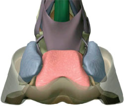 |
| Fig. 2. The structures that make up the conformation of the palmar section of the foot (frog, digital cushion, and ungual cartilages). (Courtesy of Dr. Andrew Parks.) |
Palmar Foot
The structures that comprise and form the palmar/plantar section of the foot are often the limiting factor when trying to achieve and maintain good hoof conformation or shape.5 These structures primarily include the frog, digital cushion, and ungual cartilages (Fig. 2). The conformation of the palmar/plantar section of the foot will be contingent on the accumulated mass provided by these structures. Unfortunately, attaining sufficient mass for good conformation is not always possible, because these structures are influenced by genetics (certain breeds such as the Thoroughbred will often have decreased mass), improper development or maturity of these structures, continuous repetitive overload when these structures are immature (especially seen with young horses in training), and obviously, inappropriate farrier practices. In farriery practice, it will generally be the heel area of a given foot that will need to be trimmed or manipulated to implement and attain the desired HPA, center of rotation, and hoof-capsule extension to the base of the frog.
The HPA
The HPA is our first guideline when trimming the foot. When the horse is standing still and the metacarpus/metatarsus is perpendicular to the ground and viewed from the lateral side, the HPA should form a straight line. This means that the dorsal hoof wall should be parallel to the dorsal surface of the hoof wall. The first, second, and distal phalanx along with the navicular bone comprise the bones of the digit. When the HPA is parallel, a linear line will pass through the middle of the phalanges from the metacarpal phalangeal joint to the ground. The straight alignment of the phalanges places the dorsopalmar orientation of the distal phalanx within the hoof capsule so that the solar surface of the distal phalanx assumes a similar alignment relative to the ground surface of the hoof capsule/ground. Ideally, the palmar angle of the distal phalanx should be 3-5° greater than the dorsal angle of the distal phalanx, because this allows the distal phalanx to sink in a distopalmar direction during weight bearing and use the physiology in the palmar/plantar section of the foot.3,7,12 The palmar angle of the distal phalanx is generally dependent on the conformation of the palmar/plantar section of the foot. The dorsopalmar/plantar orientation of the distal phalanx within the hoof capsule is important to avoid disproportionate load concentration on the solar surface of the hoof capsule, and it changes in the position of the ground reaction force (GRF).5 Changes in the GRF in a dorsal direction, such as the broken forward HPA, or in a palmar direction, such as in the broken back HPA, would suggest disproportionate weight bearing on the solar surface of the hoof capsule when the foot is loaded. Additional detrimental effects of either a broken back or broken forward HPA have been well documented.4,10,13 The changes in HPA are always associated with two common hoof-capsule distortions: the low or under-run heel and the upright or club foot. A broken back HPA is often caused by excessive toe length and is often combined with a low heel/under-run heel. The low heel is generally combined with a lack of soft-tissue mass in the palmar/plantar section of the foot so that the foot is unable to dissipate the energy of impact during landing. The broken back HPA also places the DIP and proximal interphalangeal joints in dorsiflexion, promotes load bearing in the heel area of the foot, and increases the stresses in the deep digital flexor tendon.
With a broken forward HPA, the heels grow long. The long heels will bypass the soft-tissue structures in the palmar/plantar section of the foot, causing the energy of impact generated during landing to be transferred directly to bone. This places the DIP and proximal interphalangeal joints in flexion and promotes load bearing on the dorsal margin of the distal phalanx.
Until relatively recently, the veterinary and farrier literature recommended that the normal hoof angle be 48-55° for the front feet and 52-60° for the rear feet. These recommendations have been shown to be erroneous, because they do not take into consideration the conformation of the horse's individual limb. Therefore, the foot is trimmed appropriately, and the hoof angle is correct for the individual horse when the dorsal hoof wall and the dorsal surface of the pastern region are aligned in parallel planes (Fig. 3, A and B).4,10 A parallel HPA is easy to access visually and can be confirmed radiographically when necessary.
The Center of Rotation
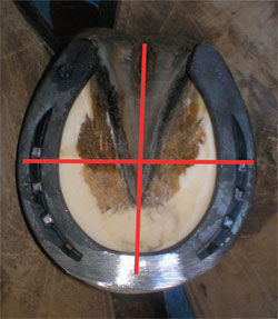
The second landmark for trimming the foot is the center of rotation. A vertical line drawn from the center of the lateral condyle of the distal second phalanx (viewed laterally) to the ground should bisect the bearing surface approximately in the middle of the foot (Fig. 4).2,4,5,10,13 This line demarcates the theoretical center of rotation of the DIP joint,and it should coincide with a line drawn across the solar surface of the foot through the middle onethird of the frog or the widest part of the foot. The widest part of the foot (center of rotation) forms a landmark on the solar surface of the foot that not only can be used as a reference point when trimming but can also be used in evaluation of foot conformation and the existing farriery that has been performed on the horse (Fig. 5). This is the point that farriers refer to as "Ducketts" bridge.a The widest part of the foot is the one point on the solar surface of the foot that remains relatively constant, regardless of the shape or length of the ground surface that is dorsal or palmar to this point. In a biomechanical sense, because the widest part of the foot is approximately under the center of rotation or biomechanical pivot point, there will be movements created on either side of the center of rotation (Fig.6). Therefore, when considering biomechanical efficiency,the distance and force on either side of the line drawn through the widest part of the foot should approximate each other (equilibrium) when the horse is standing at rest. After the trim, the ground surface of the ideal foot or good foot will be as wide as it is long, and the ground surface of the hoof capsule at the heels should not project dorsal to the base of the frog.4,5
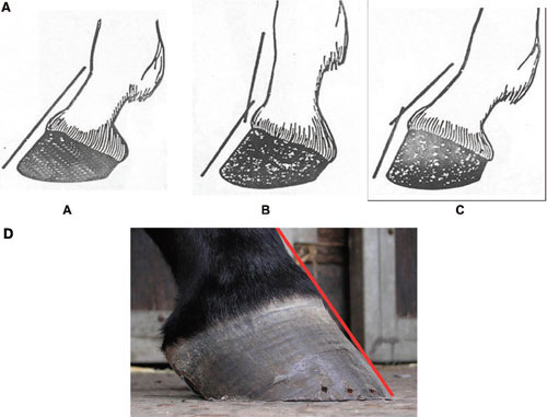 |
| Fig. 3. (A) The illustration shows a parallel HPA, (B) a broken back HPA, and (C) a broken forward hoof pastern axis. The red line in D denotes a parallel HPA. |
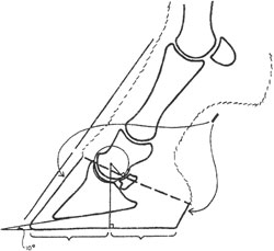 |
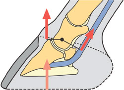
| Fig. 4. The diagrammatic drawing shows the center of rotation. |
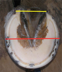 |
| Fig. 5. The widest part of the foot (red line) is located under the center of rotation. Heels are trimmed to the base of the frog (yellow line). |
 |
| Fig. 6. The illustration shows the moment or torque created on either side of the center of rotation (moment = force x length). |
Heels Extending to the Base of the Frog
The third landmark is the heels of the hoof capsule extending to the base of the frog. Present within the hoof capsule are osseous structures that will accept load through the lamellae and soft-tissue structures. These structures are designed to absorb concussion during load bearing and dissipate the energy of impact. If we consider the anatomical position of these structures within the hoof capsule, it can be noted that the distal phalanx occupies approximately two-thirds of the space, whereas the soft-tissue structures occupy one-third of the space (Fig. 7A). This is necessary for the physiologic function of the foot and for the osseous and soft tissue structures to complement each other. For this function to be effective, both the osseous and soft-tissue structures need to be enclosed within the hoof capsule in the same plane (Fig. 7B). This necessitates that the hoof wall at the heels of the hoof capsule extend to the base of the frog. Appropriate or consistent trimming of the palmar/plantar section of the foot has always presented difficulty to the clinician. Variations in hoof conformation, farrier training, empiric considerations, and owner pressure often dictate how the heels of the foot are trimmed. If the heels are left too long or are allowed to migrate dorsally toward the center of the foot, the function of the soft-tissue structures are assumed by the bones of the digit. Often, there is limited soft-tissue mass in the palmar foot or the hoof wall at the heels cannot be trimmed to the base of the frog; this necessitates that the branch of the shoe or some other form of farriery extends to the base of the frog. A discussion of farrier methods to address the palmar section of the foot is beyond the scope of this paper.
5. Method
To implement appropriate farriery (trimming and shoeing), guidelines or landmarks can be used to carefully evaluate each foot. The guidelines described are the biomechanical principles that address the HPA, center of rotation, and hoof capsule extension to the base of the frog. Good quality radiographs can be used to confirm the guidelines and provide additional parameters for farriery. Observation of the HPA in the standing horse should be the first observation or guideline in our approach to trimming the foot. To confirm that the HPA is parallel, the horse should be viewed from the side standing on a hard, level surface, and the horse must stand squarely on all four feet with the third metacarpal bone positioned vertically to the ground. If the HPA is not straight and is either broken forward or broken backward, an initial trimming plan can be generated to correct these observations.The center of rotation can also be located and marked on the outer surface of the hoof capsule. A very practical technique is to palpate the dorsal and palmar border of the second phalanx just above the coronet. One-half the distance between the borders of the second phalanx is determined, and from this point, a vertical line is dropped from the coronet to the ground (Fig. 8). This line has a good correlation with the widest part of the foot on the solar surface. Trimming begins with a line visually placed or drawn across the widest part of the foot that corresponds to a vertical line dropped from the center of the distal end of the second phalanx.4,10This line drawn across the widest part of the foot will approximate the center of rotation. Other than any loose or exfoliating horn present over the frog or the sole, the author removes no horny material from the solar surface of the foot. In most cases, any horn material that remains after the foot is cleaned with a wire brush is left intact. Excess length of the hoof wall at the toe and quarters of the hoof is determined by paring the sole/wall junction,and it is removed, being careful to leave the adjacent sole for protection. The heels of the hoof capsule are trimmed level with the toe and quarters; then,using a rasp, trimming extends to the base of the frog, when possible, with the intent being to create a solid heel base. If insufficient hoof wall is present for the end of the heel to reach the base of the frog, this distance can be lengthened by extending the branches of the shoe. The medial or lateral hoof wall can be trimmed cautiously relative to the other when changes to the lateral medial alignment of the foot are necessary. A line drawn from the initial line across the widest part of the foot to the base of the frog should approximate another line drawn from the widest part of the foot to the hoof wall at the toe. This places the center of rotation in the middle of the foot or in the middle of the shoe when shod. The foot is further shaped by removing flares from the outer surface of the hoof capsule to concentrate weight bearing on the hoof wall under the limb. This constitutes a basic fundamental trim to which a shoe can be applied to compliment the trim, protect what has been trimmed, and change the biomechanics of the foot, when necessary.
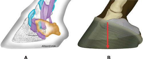 |
| Fig. 7. (A) Two-thirds of the hoof capsule is occupied with osseous tissue, and one-third of the hoof capsule is soft tissue. (B) The illustration shows osseous and soft tissue enclosed within the hoof capsule. The red arrow is the center of rotation. (Courtesy of Dr Andrew Parks.) |
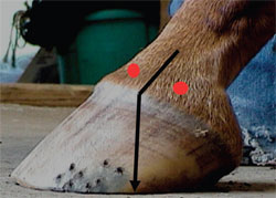 |
| Fig. 8. Red dots are the dorsal and palmar borders of the second phalanx. A vertical line drawn from a point one-half the distance between these borders to the ground will approximate the center of rotation. |
 |
| Fig. 9. The shoe is placed so that the widest part of the foot is located in the center of the shoe. Note the breakover created in the toe of the shoe to mechanically enhance breakover. |
Shoe Placement
When a shoe is placed on a horse's foot, the horse no longer stands on its feet but on its shoes instead. Therefore, the shoe is placed with biomechanical congruency so that it considers the same biomechanical landmarks used for the trim and essentially forms an extension of the trim. The shoe should be placed central to the widest part of the foot. The distance between breakover (the dorsal point of the shoe's contact with the ground) and the widest part of the foot should equal the distance from the widest part of the foot to the heel end of the shoe (Fig. 9). Breakover in the toe of the shoe can be altered when necessary by forging or by using a hand grinder. Using this technique also provides a basis in which to evaluate the existing farriery.
Radiographs
Radiography can be used as a diagnostic tool, an aid in assessing the landmarks alluded to in this paper, and a blueprint to implement these landmarks in difficult cases. Considerable information can be obtained from the image of the overall shape of the hoof capsule, the structures of the soft tissue, and the position of the distal phalanx within the hoof capsule. From the lateral medial radiographic view, one can readily assess the HPA, center of rotation, extent of the palmar hoof capsule, sole depth, angle of the palmar process of the distal phalanx,dorsal angle of the distal phalanx, breakover, and placement of the shoe.14 The dorsopalmar view will allow the clinician to evaluate the lateral medial orientation of the distal phalanx within the hoof capsule, the position of the distal phalanx relative to the ground, and the position of the distal phalanx on the joint spaces of the digit. The need to assess the position of the hoof capsule relative to the long axis of the digit is often overlooked.
6. Discussion
Adherence to the basic principles of farriery is essential for maintaining hoof health and continuous soundness. Becoming familiar with three basic landmarks will enable the veterinarian and farrier to approach trimming the equine foot in an individual, standardized, and repeatable manner. Although the guidelines or landmarks put forth in this paper are meant as a generalized guide to trimming the foot, these guidelines can also be used to recognize changes in hoof conformation that can be corrected through farriery. For example, a horse with a long toe or low/under-run heel will have a broken back HPA, whereas a horse with an upright hoof angle or club foot will have a broken forward HPA. Furthermore, if we examine the solar surface of the foot and document the center of rotation or the widest part of the foot,there will be a marked disparity in the proportion of the ground surface on either side of this landmark in either case. With the various hoof-capsule distortions,we are then left to decide how to implement the most appropriate farriery to reach the third guideline, which is to have the heels of the hoof capsule extend to the base of the frog to accommodate all the structures in the foot. Another advantage of these landmarks is the creation of a technical language that can be used to discuss farriery between the professions, and it will form the written basis for reports and records; this can be especially valuable for the new owner and farrier when included as part of the purchase examination report. Sound physiologic horseshoeing can only be achieved by a thorough knowledge of, strict adherence to, and skillful application of basic farriery principles or landmarks, such as using the HPA, center of rotation, and widest part of the frog for trimming/shoeing.Only then does the art of farriery truly approach a science. There is no question that a strong, healthy foot will enhance performance and increase the longevity of a given horse.
References and Footnote