Proper Physiological Horseshoeing: What is it? How Do We Apply It?Reprinted with permission from the American Association of Equine Practitioners. |
 |
| Figure 1. Illustration shows the relationship between the osseous and soft tissue structures within the hoof capsule (Courtesy Dr. Andrew Parks). |
Function
Classically, the horse is considered to bear weight on the wall, bars and immediately adjacent sole as well as the frog to some extent. This appears true for a horse that is wearing shoes and standing on a flat hard surface but if the ground surface is soft and deformable, more of the ground surface of the sole and frog will assume a load sharing function. Initial contact with the ground is generally made with the heels first but many horses will land flat. Toe first landing is considered to be abnormal and an indication of palmar foot pain.10 For clarity, the function or physiology of the foot will be considered during the impact and stance phase of the stride. The relationship of function to lameness is readily observed during these two phases of the stride. As the foot impacts with the ground, the physiologic process involves many structures simultaneously. The act of weight bearing is carried out by the hoof wall, adjacent sole, and partially through the frog, transferred through the lamina to the distal and second phalanx. The energy generated through impact is dissipated by the flexibility of the hoof capsule and the soft tissue structures of the palmar foot especially the digital cushion.5 Flexibility of the hoof capsule, due mainly to the properties of the hoof wall and expansion of the heels, is an interactive effort between the hoof capsule and the soft tissue structures in the palmar section of the foot. The exact mechanism by which the heels expand still remains to be documented. It is speculated that when weight is placed on the foot, the frog will flatten and spread abaxially to assist in absorbing the energy of impact. What actually appears to happen is during impact, the second phalanx presses the digital cushion down upon the frog causing it to deform.5 The digital cushion expands outwards, pressing against the ungual cartilages, spreading the quarters of the foot even more.5 Recent research has shown the bar of the heel to be located under the axial projection of the ungual cartilage and upward pressure on the bar during weight bearing causes the cartilage to be displaced abaxially.7 A series of venous plexuses located at the coronet, under the sole and adjacent to the ungual cartilages create a hemodynamic effect that contributes to the anti-concussive properties of the hoof. To further counteract the concussion associated with impact and weight bearing, the distal phalanx descends into the hoof capsule due to the elasticity of the lamina in a distopalmar direction, causing the sole to flatten; the palmar margin of the distal phalanx descends distally, pushing the navicular bone into the deep digital flexor tendon.7
Conformation of the Foot
An ideal method for a clinician to monitor and preserve foot health is through careful observation and evaluation of hoof conformation. Foot conformation can be easily monitored through serial radiographs and digital pictures. This documentation can also form part of the horse's veterinary record. Foot conformation (shape) is important because of its relationship to the foot's biomechanical function. Any changes made to the bottom of the horse's foot will affect the angulation of the hoof, the hoof-pastern axis, the ground surface of the foot and the alignment of the hoof capsule under the center of rotation.2 A review of what is considered to be good or ideal foot conformation will allow the clinician or farrier to make appropriate farriery changes when subtle changes in foot conformation are observed. The alignment of the digit, consisting of the proximal, middle and distal phalanx, should form a straight line with the solar surface of the distal phalanx having a 3-5 degree palmar angle relative to the ground.12 The hoof capsule should have a thick durable hoof wall, greater than 15 millimeters of sole depth and a well-defined frog where the width approximates the length. The ground surface of the hoof capsule should be basically as wide as it is long. The dorsal surface of the pastern and the dorsal hoof wall should be parallel. This is termed the hoof-pastern axis. The coronet should have a gradual uniform slope from the toe to the heel. The literature states that the angle of the dorsal hoof wall and the angle of the heel should correspond yet this is seldom the case. The hoof wall at the toe is mature, stiff fixed horn whereas the hoof wall at the heel is immature, thin and flexible to allow for expansion of the palmar hoof capsule. During load bearing, whether shod or barefoot, the heels move abaxially either against the shoe or to a lesser extent against the ground when barefoot leading to wear at the heels when compared to the toe. This continual abrasion at the heels leads to wear and results in the heel angle being generally lower than the toe. A marked decrease in heel angle relative to the toe angle is considered a low or under run heel. Finally, the ideal foot has a well-defined heel base consisting of good hoof wall, a solid angle of the sole and straight bars (Fig. 2).
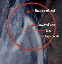 |
| Figure 2. Heel base consists of the buttress of the heel, angle of the sole, bar and hoof wall. |
For simplification in allowing veterinarians and farriers to observe subtle changes in hoof conformation, three visual references will be used; the hoof-pastern axis, the widest part of the foot and the base of the frog. The hoof-pastern axis is correct for an individual horse when the dorsal hoof wall and the dorsal surface of the pastern region are aligned in parallel planes11 (Fig. 3).This is best observed with the horse standing squarely on a flat hard level surface with the third metacarpal bones positioned vertically relative to the ground. Changes in hoof-pastern axis such as a broken back or broken forward hoof-pastern axis are always a reflection of hoof conformation. The other significant guideline that can be used to evaluate hoof conformation is the widest part of the foot. In the "ideal" foot, a vertical line drawn from the center of the lateral condyle of the distal second phalanx (viewed laterally) to the ground should bisect the middle of the bearing surface of the foot.12 This line marks the theoretical center of rotation of the distal interphalangeal joint and should coincide with a line drawn across the widest part of the foot. The widest part of the foot or center of rotation forms a landmark on the solar surface of the foot that can be used to access foot conformation and also will be used as a reference point when trimming. Thirdly, to accommodate or enclose both the distal phalanx and the soft tissue structures palmar/plantar to the bone, the heels of the hoof capsule should extend to the base of the frog (Fig. 4).
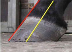 |
| Figure 3. Parallel hoof-pastern axis. Yellow depicts digital alignment. |
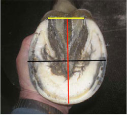 |
| Figure 4. Black line shows the widest part of the foot . Yellow line denotes the base of the frog. Corresponding red line from the base of the frog to the toe shows a well conformed foot is as wide as it is long. |
Proper Physiological Horseshoeing
Conventional wisdom on trimming and shoeing horses is that each case should be regarded as an individual. Central to our current knowledge of farriery is the interaction of the structures of the hoof and the manner in which the foot loads and the surface upon which the horse is asked to perform. Our approach to proper physiological horseshoeing should address three parameters: the visual structures of the hoof complex, function of the distal interphalangeal joint and the biomechanical forces applied to a given foot. Dorsal palmar and medial lateral orientations are dictated by form and placement of the distal phalanx within the hoof complex. A decrease in the angle between the palmar margin of the distal phalanx and the ground surface results in greater loading of the navicular bone, while differences between the toe angle/heel angles has not been correlated to increase in loading of the navicular apparatus.
How Do We Apply It?
To implement basic trimming and shoeing, guidelines are established using careful evaluation of the foot along with good quality radiographs when necessary. Trimming and shoeing techniques are applied to the foot using the biomechanical principles that address the hoof-pastern axis, the center of articulation and extending the heels of the hoof capsule to the base of the frog. Prior to trimming the foot, the hoof pastern axis is evaluated with the horse standing on a firm, flat surface such that the third metacarpal bone is perpendicular with the ground. A straight or parallel hoof pastern axis indicates that the distal phalanx is positioned within the hoof capsule such that load is accepted on the entire solar surface of the hoof. With a broken back hoof pastern axis, load will be concentrated in the palmar section of the foot and with a broken forward hoof pastern axis; the load will be borne in the dorsal or toe section of the foot. Trimming the foot begins with a line visualized or drawn across the widest part of the foot. This line corresponds to a vertical line dropped from the center of the distal end of the second phalanx to the ground.13 This line drawn across the widest part of the foot will approximate the center of articulation. Other than excess exfoliating horn material, no horn is removed from the sole surface or frog. Excess length of the hoof wall at the toe of the hoof is determined at the sole/wall junction and removed, being careful to leave the adjacent sole for protection. Next, the heels are trimmed with a rasp to the base of the frog when possible with the intent being to create a solid heel base and including both the osseous and soft tissue structures within the hoof capsule. If insufficient hoof wall is present for the end of the heels of the hoof capsule to reach the base of the frog, this distance can be lengthened with the shoe. The medial or lateral wall can be lowered cautiously relative to the other when changes to the lateral medial alignment of the foot are necessary. A line drawn from the existing line across the widest part of the foot to the base of the frog should be equal to another line drawn from the widest part of the foot to the toe. Excess length of hoof wall can be removed by backing up the dorsal hoof with a rasp from the outer surface of the hoof capsule to create the desired distance to approximate the measurement from the middle of the foot to the heels. This places the center of articulation in the middle of the foot or in the middle of the shoe when shod with approximately equal distances on either side of the widest part of the foot. This creates a foot that is basically as wide as it is long which is thought to be biomechanically efficient. The foot is shaped by removing excess flares from the outer surface of the hoof capsule to concentrate weight bearing on the hoof wall. This constitutes a basic fundamental trim to which a shoe can be applied to compliment the trim, protect what has been trimmed and to change the biomechanics of the foot further when necessary. When the shoe is placed on the foot, the line drawn across the widest part of the foot will be in the middle or center of the shoe. The shoe should be as light as possible, steel or aluminum, with a wide web width (5/8-3/4 inch) attached with as few nails as possible of the smallest size.
Hoof Capsule Distortions
Broken Back Hoof Pastern-Axis
A broken back hoof-pastern axis will be a reflection of a hoof capsule where the angle of the dorsal hoof wall is lower than the angle of the dorsal pastern (long toe/low or under run heel conformation). This type of foot configuration is so common in equine practice that it is thought to be normal. In one study of foot related lameness it was found in 77% of the horses2 and in another study of normal performance horses this condition was found in 52% of the horses.9 A low hoof angle causes coffin joint dorsiflexion, concentrates weight bearing on the palmar section of the foot and increases strain on the DDF tendon. This excess load in turn, may cause increased stresses on the navicular apparatus and the soft tissue structures associated with the navicular bone. Excessive toe length is thought to delay the speed of breakover. If, as a result of a low hoof angle, the horse begins to experience pain in the heel region, it will land toe first; this may lead to subsolar bruising. This abnormal hoof conformation may contribute to palmar foot pain, chronic heel bruising, coffin joint synovitis, quarter and heel cracks and interference problems. There is also experimental evidence that a low hoof angle will compromise circulation in the heel area of the foot.12 It may be helpful to make a distinction between a low heel and an under run heel. In the case of a low heel, the angle of the heel will be markedly lower than the angle of the dorsal hoof wall; however, the structure of the heel is relatively good, in that the buttress, angle of the sole and bars are intact forming a base. In the under run heel, the structure of the heels is compromised such that the hoof wall at the heels is thin, separated and rolled under in a axial direction, the angle of the sole is missing, the bars are destroyed and the heel-ground contact does not reach the base of the frog. As low or under run heels progress, this condition can be readily observed both visually and radiographically, where the angle that the hoof capsule or the distal phalanx forms with the ground will be lower palmarly/plantarly than it is dorsally. A common error in routine farriery is not continually moving the ground surface of the hoof wall at the heels to or toward the base of the frog. A negative palmar angle, as noted radiographically, basically means that the soft tissue structures (frog, digital cushion) have decreased in mass usually due to damage or they have prolapsed palmarly (Fig. 5). This type of hoof conformation alters the mechanics of the foot as the compromised heels lose both the ability to accept weight and to dissipate the energy of impact.
 |
| Figure 5. Negative hoof pastern axis which alters digital alignment so weight bearing is moved palmarly. |
Broken Forward Hoof-Pastern Axis
A broken forward hoof-pastern axis will be a reflection of a hoof capsule where the angle of the dorsal hoof wall is higher than the angle of the dorsal pastern (upright or club foot conformation). A high hoof angle leads to coffin joint flexion, promotes toe first landing and increases pressure in the dorsal section of the foot. Poor performance and injuries associated with a high hoof angle are thought to include coffin joint inflammation, due to abnormal loading of the joint, sole bruising, and increased strain on the suspensory ligaments of the navicular bone.
High hoof angles without phalangeal misalignment can be improved by gradually lowering the heels in a tapered fashion from the apex of the frog to the heels. This increases the ground surface of the foot and attempts to re-establish weight bearing on the entire solar surface of the foot. Breakover is moved palmarly at the same time to compensate for any increased tension in the DDFT created by lowering the heels.
An extremely high hoof angle with concurrent phalangeal misalignment is often classified as a flexural deformity or "club foot". This broken forward hoof-pastern axis or flexural deformity is created by a shortened musculotendonous unit (deep digital flexor tendon and associated muscle bellies) causing the distal interphalangeal joint (DIP) to be drawn into a flexed position. Flexural deformities have been reported as a cause of decreased athletic performance and chronic low grade lameness in the mature horse.13,14 Hoof abnormalities associated with club foot conformation are thin flat soles, poor hoof wall consistency, toe cracks, hoof wall separations and "white line disease". Flexural deformities that result in a marked broken forward hoof-pastern axis have also been associated with chronic distal interphalangeal joint inflammation.14 Generally, flexural deformities are diagnosed and treated while the horse is immature. However, mild flexural deformities may be ignored or treated improperly as a foal. When these animals enter training, mild flexural deformities can be exacerbated by the type and the amount of exercise, the ground surface, by inappropriate farrier care, such as improper or infrequent shoeing, or by some type of underlying pathology. The treatment of advanced flexural deformities is beyond the scope of this paper.
 |
| Figure 6. Sheared heel conformation. Medial heel is displaced proximally when compared to the lateral side. Note flare on lateral side. |
Sheared Heels
Another common variation in hoof conformation is sheared heels. A sheared heel is a hoof capsule distortion resulting from displacement of one heel bulb proximally relative to the adjacent heel bulb.15 (Fig. 6) This disparity between the lateral and medial heel bulb is generally 0.5 centimeters or more. When the weight of the horse is not distributed uniformly over the entire hoof during the landing phase of the stride, one focal area of the foot, usually a heel or heel and accompanying quarter, receives a disproportionate amount of the total force during impact. This resultant force leads to a remodeling of the affected heel bulb. Although a number of sound horses have this type of hoof capsule distortion, lameness is often attributed to this condition. This continual disproportionate loading and increased compressive stresses on one heel predisposes the foot to hoof capsule distortion, subsolar bruising, corns, quarter and heel cracks, fracture of the bar deep fissures within the base of the frog and thrush in narrow frogs. This type of conformation is readily observed by picking up the foot and noting the relative distances measured from the heel of the hoof capsule to the hairline at the bulbs of the heels between the lateral and medial heel.
Inappropriate lateral medial orientation (balance) of the foot or a landing pattern where the foot does not land flat has always been associated with improper trimming. However, there appears to be more of a correlation between limb conformation from the carpus distally that changes the flight of the limb and ultimately the manner in which the foot lands.16 Furthermore, there appears to be a correlation between an offset distal phalanx and sheared heels. Most commonly the distal phalanx is offset laterally within the hoof capsule rather than directly under the first and second phalanges with the ensuing concussive forces causing the medial heel to displace. Farriery practices have always advocated trimming the horses with sheared heels so the ground surface of the foot is lower on the opposite side from the one that is displaced proximally. Intuitively, if the heel is longer on the displaced side (measured ground surface to hairline), it is reasonable to lower the affected side. This in fact changes the landing pattern of the horses when it is observed in motion following this type of trim. Therefore, when this type of conformation becomes apparent, lowering the affected side and fitting a symmetrical shoe is a reasonable approach.
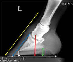 |
| Figure 7. Lateral radiograph used to illustrate hoof-pastern axis (yellow), center of articulation (red), sole depth (black), and palmar angle of distal phalanx (green), breakover (blue) and the placement of the shoe (white). |
Radiographs
Radiography can be utilized as both a diagnostic tool and as an aid in assessing all structures of the foot when necessary. The radiograph can serve as a blueprint for veterinarians and farriers which can be used as a guideline for applying farriery. Considerable information can be obtained from the image of the overall shape of the hoof capsule, the soft tissue structures, and the position of the distal phalanx within the hoof capsule. It should be remembered that lack of performance or many subtle lameness cases localized to the foot are caused by hoof capsule distortions, poor foot conformation, improper landing patterns, and soft tissue damage resulting in inappropriate biomechanical stresses being placed on the navicular complex and the distal interphalangeal joint. The clinician will be able to use the lateral to medial and dorsopalmar views of the foot as a precise guide to implement basic or therapeutic trimming and shoeing (Fig. 7). From the lateral medial view we can readily measure the hoof pastern axis, the center of articulation, sole depth, the palmar angle of the distal phalanx, breakover, and the placement of the shoe. When the solar plane of the distal phalanx shows a negative palmar angle, we can also access the need for heel elevation and the amount to apply. The dorsopalmar view will allow the clinician to evaluate the lateral medial orientation of the distal phalanx within the hoof capsule, the position of the distal phalanx relative to the ground, the effect of the position of the distal phalanx on the joint spaces of the digit and then make the appropriate shoeing changes. The need to access the position of the hoof capsule relative to the long axis of the digit is often overlooked.
Applying Proper Physiological Horseshoeing to the Performance Horse
Shoes are essential for the performance horse not only to protect the hoof but also to preserve the hoof complex and the structures contained within the hoof capsule during the rigors of competition. Enormous differences exist between different equestrian disciplines, the different breeds of horses used in these disciplines, the ground surface on which they perform along with different shoeing materials and shoeing styles. But it should be remembered that all horses have the same basic anatomy and physiology and sound farriery principles as outlined previously can be applied regardless of the breed or the discipline. Special attention is paid to the mass and heel base present in the palmar/plantar section of the foot as the shoe protects the toe of the hoof from wear but not the heels. During weight bearing, the hoof wall at the heels move against the shoe leading to wear. This wear is exaggerated during prolonged competition where the feet are subjected to repetitive high impact stresses such as those encountered during racing, jumping, and eventing. Hoof mass is very important in show jumpers as they continually stress the flexor structures within the hoof and the podotrochlear apparatus. It is important to preserve the bars and recruit them into weight bearing. A subtle change in heel angle is the first indication of a change in foot conformation. Employing the use of leather pads and deformable packing materials such as the pour-in padsa will play an important role in preventing excessive wear at the heels and also absorbing concussion especially in the horse with an upright or club foot.
Wide web aluminum or steel shoes creased at the toe but not at the heels (to accommodate stud holes if necessary) are used in most sport horses. The use of toe or side clips stabilize the shoes especially when pads and studs are used, thus relieving stress on the nails and allowing the use of fewer nails. Strict attention should be paid to hoof wall length and breakover in all sport horses. Moving breakover palmarly/plantarly can be accomplished in a variety of ways such as rockering the toe of the shoe or creating a rolled toe in the shoe using a hand grinder where the breakover begins at the toe quarters (Fig. 8). Another method to facilitate breakover is through the use of half round shoes. Moving breakover palmarly/plantarly will decrease the moment applied to the distal interphalangeal joint and appears to decrease the maximum tension in the deep digital flexor tendon which occurs towards the end of the stance phase at the beginning of breakover.17 Bar shoes, especially egg bar shoes, are very popular in sport horses. Bar shoes if fitted properly will stabilize a weak hoof capsule and increase the ground surface in all applications. Caution should be used when applying egg bar shoes to horses with damaged or under run heels. The shoe is usually fitted long extending to the bulbs of the heels with the misguided thought that they will support the heels. In reality, egg bar shoes will create a moment exerted on the damaged heels creating pressure and consequently stopping growth. If bar shoes are indicated or desired, the author prefers straight bar shoes that when fitted properly, follow the contour of the hoof capsule and extend a few millimeters beyond the base of the frog (Fig. 9). Egg bar shoes or any bar shoes will increase concussion forces on a hard surface but may decrease these forces on a deformable surface by providing a flotation effect where as the increased ground surface of the bar does not allow the heel to readily sink into the ground.18
 |
| Figure 8. Picture shows breakover cut into the toe of an aluminum shoe extending from the toe quarters dorsally to the periphery. |
 |
| Figure 9. Straight bar shoe with breakover moved palmarly and impression material placed between the branches of the shoe to increase surface area. |
Shoes in and of themselves provide traction. Traction is enhanced when a shoe with a crease is used. Shoes are often combined with various traction devices such as studs, chalks and borium that are placed in or on the shoe to further aid traction, enhance performance and provide safety to horse and rider. The stud holes should always be drilled at the end of the quarter crease rather than at the end of the shoe branch. This will eliminate the stud's lever arm effect on the digital joints during sharp turns. Screw-in studs have the benefit of being able to be removed when the horse is not performing.
Conclusions
Adherence to the basic principles of proper physiological horseshoeing is essential for maintaining hoof health and continuous soundness. Most horses do not require special trimming or shoeing techniques. Becoming familiar with a few basic concepts can help the veterinarian recognize when changes in trimming and/or shoeing might be expected to help the performance of a sound horse, or might help to restore the performance of one that is lame. Sound physiologic horseshoeing can only be achieved by a thorough knowledge of, strict adherence to, and the skillful application of basic farriery principles such as utilizing the hoof pastern axis, the center of articulation and trimming/shoeing to the widest part of the frog. Only then does the art of farriery truly approach becoming a science. There is no question that a strong healthy foot will ensure comfort, enhance performance, and increase the longevity of the horse.
References