Farriery for the foal: A review part 1: Basic trimmingReprinted with permission from Equine Veterinary Education (EVE). |
| ||
| ||
 | ||
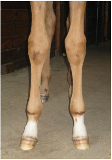
| Fig 3: The width is wider at the coronet than at the ground surface of the foot in this 6-week-old foal. Also note the pointed toe. |
Trimming the foal
Birth to one month
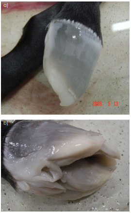
At birth, the foal’s hoof is enveloped in a gelatinous perioplic membrane (eponychium) which reduces the risk of trauma to the mare’s reproductive tract during gestation and parturition(Fig 1). Shortly after birth, with the first steps of life, the perioplic membrane on the solar surface of the foot wears,dehydrates and retracts proximally on the hoof wall and ultimately creates a sulcus of varying depths just distal to the coronet the coronary band (Fig 2a and b). This depression,termed the sub-coronary groove, is considered normal and grows distally towards the ground. In lay terms it is often referred to as a ‘milk’ foot as it appears at birth and generally has grown out over the next 4–6 months by weaning. It has the potential to cause a defect or separation in the sole wall junction (white line) when it approaches the ground surface of the foot if the toe length is allowed to grow excessively long. The remnants of the sub-coronary groove create leverage at the toe if not trimmed appropriately and this force is responsible for a bending in the dorsal hoof wall or separation at the sole wall junction in the toe. The foal’s foot at this time is generally tapered, being wider at the coronet and becoming narrower distally at the ground surface (Fig 3). A foal`s foot does not only grow in a distal direction, but it also expands as it develops. As the foal’s feet are tapered, expansion occurs proximally and as the ground surface of the distal hoof is relatively small, the weight-bearing area is positioned in the dorsal section of the foot. Exercise and appropriate trimming will enlarge the area on the ground surface of the foot and move it in a palmar/plantar direction. The pointed or tapered appearance will gradually disappear in the first few months of life with appropriate trimming. In foals with acceptable limb conformation there is little need for trimming during the first month of life.
One month
Foals should be presented to the farrier at one month of age for trimming. Prior to the first trimming, basic limb and foot manipulations by trained farm personnel should have the foal accustomed to the positioning used by the farrier. Trimming should be a pleasant experience for the foal and will act asa form of imprinting if started in a gentle manner from the beginning. The farrier should be patient, perform the farriery gently and efficiently and not fight the foal. An experienced handler that is gentle but firm is essential. The use of a nose twitch or chemical restraint should be discouraged. If restraint is necessary, the author will use a piece of bailing twine that is threaded through the rings of the halter and placed under the upper lip of the foal. Mild pressure or gentle tugs (never harsh) are applied to the string if necessary while the trimming is taking place. Generally, it is only necessary to use this method during the initial trimming session. The foal is always trimmed in a stall placed alongside the mare that is positioned against the wall and backed into a corner. The outer side of the foal is trimmed; then the positioning of the mare is reversed, and the other side of the foal is trimmed.
All that is generally necessary at 1 month of age is to square the toe of the hoof with a rasp to remove the tapered or pointed contour of the dorsal distal hoof wall perimeter and encourage the foal to break over in the centre of the foot. At this age, due to the pointed toe, the foal may break over to either the outside or inside of the toe(Fig 3). If the frog has receded below the level of the hoof wall, the heels should be rasped lightly using the smooth side of the rasp until the hoof wall and the frog are on the same plane. Any sharp edges are removed from the perimeter of the hoof capsule using the rasp at an angle. As will be discussed below, the use of a hoof knife or hoof nippers is discouraged when trimming foals at any age.
Two months onward
During these first few months of life, attention should be directed towards the structural integrity of the hoof capsule(foot mass/density) rather than to cosmetics. The important concerns are to promote the growth of a thick, durable hoof wall, to ensure maximum sole thickness in order to protect the vulnerable sole wall junction, the soft tissue structure and developing distal phalanx and finally to develop the structures in the palmar/plantar section of the foot. Promoting the structural mass of the foot in a foal (defined as a strong hoof wall, adequate sole depth and a solid heel base) is vital for hoof capsule development and future soundness. It is the author’s opinion that a hoof pick, wire brush and a rasp are the only tools necessary to trim foals that are kept on a month to 5 weeks trimming schedule. Furthermore, if the foal has adequate exercise combined with a consistent trimming schedule, there is generally minimal hoof growth which makes the use of a hoof knife and hoof nippers unnecessary.The goal is to not have the foal walk exclusively on the hoof wall but rather load all the structures on the solar surface of the foot; having the foot ‘load-sharing’ causes stimulation,adaptation and promotes growth. Foals that are trimmed frequently and have a lot of horn removed tend to develop weak fragile hoof capsules (O’Grady 2017).
The recommended technique of trimming foals used by the author may differ from traditional farriery (O’Grady 2008,2017). Dirt and debris are removed from the sole and sulci of the frog using a hoof pick. The solar surface of the foot is then cleaned vigorously using a wire brush to remove any loose exfoliating horn. Any loose or exfoliating tags of horn are removed from the frog with a hoof knife if necessary.Otherwise, the ground surface of the foot and the frog are left untouched which affords the foal ample protection on the ground surface of the foot. Exfoliating horn from the sole will be continuously shed through the abrasive friction with the ground as the foal exercises. The sole of a foal is relatively thin (which can be demonstrated by showing deformation when using thumb pressure or small hoof testers is applied to the sole) and needs to develop as much thickness as possible in order to protect the immature developing structures within the capsule. Removing excess sole with a hoof knife appears to be the primary cause of sole bruising in foals and may potentially lead to flexural deformities because of the pain response (Hunt 2011; O’Grady 2012). The health of the foot throughout the animal’s life is based on developing good solid heel structures. The heel base includes the hoof wall at the heel, the bars, angle of the sole, a thick digital cushion and a wide healthy frog. The bars should not be removed as they are needed for strength and to stabilise the palmar section of the hoof capsule.
| ||
|
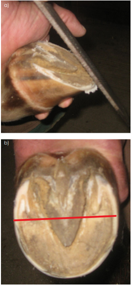
After cleaning the foot, the heels of the hoof capsule are rasped gently from side to side until the rasp just contacts the frog. The hoof wall at the heels will now be on the same horizontal plane with the frog and the heels of the hoof capsule will generally extend to the widest part of the frog.When the heels are trimmed in this manner, the frog will function as an expansion joint to keep the heels wide apart and share some of the weight bearing function. The excess hoof wall at the toe and quarters is then reduced as necessary using a rasp placed at a 90° angle just dorsal to the sole wall junction (white line) at the inner part of the stratum medium of the hoof wall (Fig 4a). When the desired amount of hoof wall is removed, the outer sharp edge around the perimeter of the foot that is formed by the angulation of the rasp is removed by running smooth side of the rasp around the perimeter of the hoof in a horizontal direction thus creating a rounded edge (Fig 4b). This round edge will help to prevent cracks and chips in the hoof wall.As the foal grows and develops, the foot assumes the same growth pattern as the adult horse which follows the angulation of the horn tubules in the hoof capsule; i.e. the heels and toe grow forward relative to the centre of rotation.Therefore, the same guidelines for trimming can be applied to the juvenile horse; using the widest part of the foot,trimming the heels to the base of the frog and creating approximate proportions on either side of a line visualised across the widest part off the foot (O’Grady 2008, 2009,2017). There is a tendency not to trim the heels appropriately in a foal and the author believes this practice detracts from the proper development of the palmar section of the foot(Fig 5a and b). The farrier should abandon the concept of lowering the heels in the foal and perhaps consider the notion of increasing the ground surface. The method of using the rasp on an angle leaves the hoof wall and the adjacent sole on the same plane allowing both structures to share the bulk of the weight when the animal moves. It also appears to stimulate the horn to grow thicker and stronger (O’Grady 2008, 2017).
Foals given sufficient exercise do not grow an excessive amount of hoof wall in the first few months of life and our ability to influence the foot/limb by excessive trimming on one side of the foot in the horizontal plane is limited and should be discouraged. If it becomes necessary to lower one side of the foot past the point of being level due to a developing hoof capsule distortion or in an attempt to affect landing, it should not be lowered any more than few millimetres at one time. Trimming the foot at 2-week intervals may be useful when trying to change the medial lateral orientation of the foot or when trying to increase the ground surface on one side of the foot.
The traditional theory of lowering the lateral side of the foot on a foal that stands toed-out or lowering the medial side of the foot on a toed-in foal is unrealistic. In fact, it maybe detrimental as the cause of the foal having a toe-in or toe-out stance generally reflects the conformation of the limb and is rarely limited to the foot (Hunt and Baker 2017). A deviation is generally found in the axial alignment of the limb above the foot such as a rotational deformity of the limb distal to the carpus or the fetlock; therefore, when one side of the foot is trimmed excessively, the cosmetic appearance may be improved temporarily but over time this practice will lead to distortion of the hoof capsule (compressed growth rings and the hoof wall rolling axially on the lowered side)resulting from an unequal load on the foot. This practice will also place excessive and unequal forces on the physes and joint on the side that is being trimmed excessively. The effects of over trimming can be observed radiographically a few days after the trim (Hunt and Baker 2017; O’Grady 2017).
Rotational deformities are very common in foals and should not be considered abnormal. For example, a narrow chest coupled with short neck and relatively long forelimbs will cause many foals to adopt a base-wide stance in front in order to graze comfortably which will often be accompanied by outward rotation of the entire limb. When viewed from the frontal plane, the entire limb will be rotated outwards,but the axial alignment of the limb will be relatively straight(Fig 6). This stance can be considered normal in foals as it confers a higher degree of stability and is gradually modified as the transverse diameters of the upper body increase with growth. As the foal moves, it is quite noticeable that the lateral side of the hoof wall strikes the ground initially because of the flight pattern caused by the rotated position of the limb. These foals should be trimmed flat or level and not have their feet lowered on the outside wall which is the traditional practice. A base-wide stance in a 3- to 4-month old foal will often result in asymmetric hoof capsules noted in the frontal plane. The medial side of the hoof capsule will be slightly lower than the lateral aspect due to the landing pattern of the foot. If this stance is not recognised as physiological for the age and an attempt is made to‘correct’ it by lowering the lateral wall, there may be a risk of creating an angular limb deformity where none existed previously. In cases where the medial heel bulb has been displaced proximally because of the asymmetrical landing pattern, although it may seem counterintuitive, the medial hoof wall is trimmed slightly more than the lateral side to create additional ground surface under the medial wall. If the medial hoof wall begins to roll under axially, the author has seen improvement by placing a small composite extension on the outer hoof wall that is used to create additional ground surface and address this hoof capsule distortion. Therapeutic trimming does not offer favourable results in the mal-positioned limb, as this deformity is corrected through growth. As the musculature of the chest increases, the chest widens and the elbows are pushed outward, which rotates the limbs inward.
|
|
| ||
|
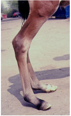
Flexor tendon flaccidity
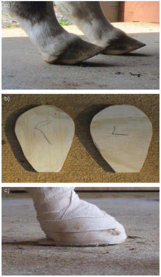
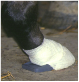
Excessive (flexor tendon) laxity in the newborn foal most commonly affects the fetlocks of the hind limbs whereas the forelimbs generally involve the fetlocks and carpi; many foals will improve spontaneously with good husbandry (as described below) and no other treatment and will have a good prognosis. This condition is often seen in premature,dysmature or septic foals (Coleman and Whitfield-Cargile 2017). When seen in the forelimbs, there is a ‘bowed’ appearance to the limb when viewed from the side from a laxity of the flexor apparatus of the entire limb. The carpus and fetlock are hyper extended with the palmar surface of the pastern and fetlock on or close to the ground. There maybe sub luxation of the distal interphalangeal joint (DIPJ)associated with deep digital flexor tendon laxity allowing the toe of the foot to elevate off the ground. In hind limb laxity,the DIPJ is almost always involved along with laxity noted in the pastern and fetlock (Fig 7). Initial treatment is aimed at protecting the soft tissues of the heels without over supporting the fetlock which will further promote the laxity. This can be accomplished by applying a self-adhesive pad (Equate®Moleskin Padding) cut in the shape of the heel bulbs. The condition tends to be self-limiting within a few days after birth as the foal gains strength and is allowed moderate exercise.However, the tendon laxity often persists and it is not uncommon to see a foal that still has digital hyper extension at 3–4 weeks of age. Treatment is sequential depending on the severity of the tendon laxity and the initial response of the foal to treatment. Therapy begins with controlled exercise allowing the foal access to a small area with firm footing for 1 h, 1–2 times daily. If there is no response by the third day post-partum, the author will place the foot on a small piece of ¼ inch plywood and trace the foot leaving 2–3 cm of extension beyond the heels. The plywood is attached to the foot using a soft kling gauze to envelop the foot and then securing the extension to the foot using 2-inch elastic tape applied in a figure of 8 technique (Fig 8a,b and c). The exercise schedule is continued and the bandages securing the extensions are reset as necessary. The laxity will generally resolve in 7–10 days and exercise can gradually be increased. When presented with an older foal, even though the toe is off the ground, the toe length of the hoof capsule should be reduced vertically or from the outer hoof wall so no leverage is applied to the toe when the ground surface of the hoof capsule is weight bearing following the application of an extension. The heels can be rasped gently from the middle of the foot palmarly/plantarly to create additional ground surface in that section of the foot; some form of a palmar/plantar extension should then be applied which extends approximately 3–4 cm beyond the bulbs of the heels to relieve the biomechanical instability of the digit (Fig 9). Acuff-type extension shoe is commercially available or a thin aluminium plate can be fabricated as an extension shoe with the aluminium bent at the toe to align with the dorsal hoof wall to hold it in place. The author feels that either type of extension shoe should be attached with the hoof enveloped in gauze and attached with elastic tape applied in a figure of 8 pattern rather than a composite if the foal is less than 3 weeks of age. This manner of attachment avoids excessive heat being applied to the fragile hoof capsule when the composite cures and the detrimental consequences that may follow. Taping the extension in place also prevents contracture of the hoof capsule which occurs at the heels when an acrylic composite is used. Heel extensions should extend beyond the heel bulbs or further; if not of sufficient length, the extension will serve as a fulcrum and worsen the subluxation of the distal interphalangeal joint and metatarsophalangeal joint. Regardless of the method of application, the extensions should be changed at 7–10-day intervals or sooner if indicated by the extension shifting.Bandaging the limb is contraindicated as the counterpressure will further weaken the flexor tendons and promote laxity
|
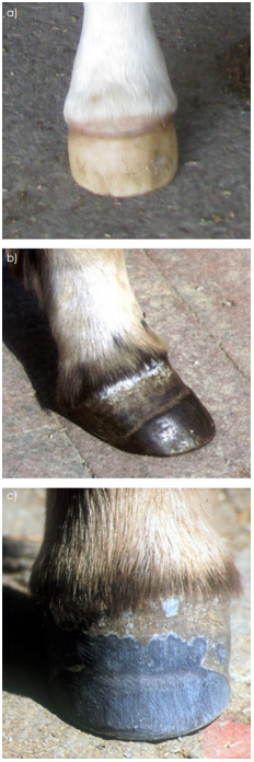
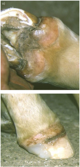
Care should be taken to maintain the condition of the feet while the tendon laxity is being addressed and long-term maintenance of the feet may be necessary. During treatment, the heels become distorted and the hoof wall growth is oriented dorsally which requires gradual re-shaping once the tendon laxity is resolved. The heels of the hoof capsule should be trimmed to the level of normal tubular alignment if possible and the heels of the hoof capsule should be on the same plane as the frog. If the foal had been allowed to walk on the bulbs of the heels for an extended period of time, there may be a demarcation or groove between the coronet and the heels of the hoof capsule (Fig 10a). The author has been successful improving horn tubular growth and alignment by filling the groove with an acrylic composite (Fig 10b). The toe length should be trimmed or reduced as necessary. This process may require 3–4 months to accomplish but over time a normal foot should and can be the result.
Conclusion
Routine hoof care in the first few months of life should never be taken lightly. The importance of good farriery in the foal plays a vital role in both the development of the hoof and the conformation of the limb. Management of hoof capsules and limbs during this juvenile period will often impact the success of the foal as a sales yearling or mature sound athlete. Foal trimming should always be based on good basic farriery principles and the appropriate biomechanics.Hoof care in the foal should always be a joint venture between the veterinarian and the farrier. The importance of maintaining a good veterinarian-farrier relationship should be emphasised; the farrier is responsible for basic trimming with veterinary oversight and if orthopaedic disorders develop, the farrier will have significant input with therapeutic farriery. A sound foot care programme is time-consuming whereas assembly-line trimming is quick and easy, but the former is much more beneficial with a better outcome. Flexural and angular limb deformities in foals will be covered in part 2 of this review.
Conflict of Interest
No conflicts of interest have been declared.
Declaration of Ethics
Not applicable.