Farriery for the Young HorseReprinted with permission from the American Association of Equine Practitioners. |
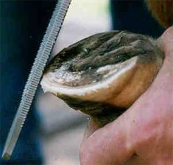 |
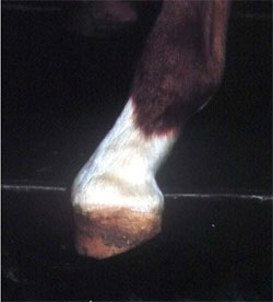
| Fig. 1A. Hoof wall trimmed on an angle dorsal to the sole wall junction. |
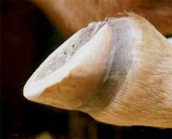 |
| Fig. 1B. Perimeter of hoof wall has a rounded edge following trim. |
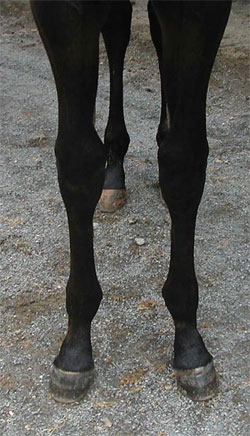 |
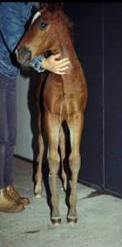
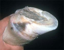
| Fig. 2. Rotational deformity in a foal. |
The method of trimming foals used by the author may differ from traditional farriery. Dirt and debris is removed from the foot using a hoof pick. The bottom of the foot is then cleaned vigorously using a wire brush to remove any loose exfoliating horn. Otherwise, the ground surface of the foot and the frog are left untouched. This affords the foal ample protection on the ground surface of the foot. Exfoliating horn from the sole will be continuously shed through an abrasive mechanism with the ground as the foal exercises. The sole in the foot of a foal is extremely thin and as much protection as possible is necessary to protect the immature developing structures above. Removing excess sole with a hoof knife is a primary cause of sole bruising in foals and often leads to flexural deformities as a result of the pain response. The health of the foot throughout the animal's life is based on a good solid heel area. The heel base includes the hoof wall at the heel, the bars and a nice wide frog. The bars are needed to stabilize the hoof capsule and are never removed. The heels are rasped gently from side to side until the rasp just comes in contact with the frog. The hoof wall at the heels will now be on the same plane with the frog. The excess hoof wall at the toe and quarters is then lowered as necessary using a rasp placed at a 90° angle just in front of the sole wall junction (white line) (Fig. 1). When the desired amount of hoof wall is removed, the outer sharp edge of the angle is removed by running the rasp around the perimeter of the hoof thus creating a nice rounded edge (Fig. 2). This will help to prevent cracks and chips in the hoof wall. The method of using the rasp on an angle leaves the hoof wall and the adjacent sole on the same plane allowing both structures to share the bulk of the weight when the animal moves. This appears to stimulate the horn to grow thicker and stronger. Foals do not grow an excessive amount of hoof wall in the first few months of life and our ability to influence the foot/limb by excessive trimming on one side of the foot in the horizontal plane is limited. If it becomes necessary to lower one side of the foot past the point of being level due to a developing hoof capsule distortion or to affect landing, it should not be any more than 2-3 millimeters at one time. Trimming at two-week intervals may be useful in this situation.
The traditional theory of lowering the lateral side of the foot on a foal that stands toed-out and lowering the medial side of the foot on a toed-in foal is inaccurate. It in fact may be harmful rather than beneficial. The cause of the foal having a toe- in or toe- out stance is rarely in the foot. The problem is generally found in the axial alignment of the limb above the foot, therefore, when one side of the foot is lowered excessively, the cosmetic appearance may be improved but over time leads to distortion of the hoof capsule. This practice will place stresses on the physis and overload the joints on the side that is being lowered. This can be shown radiographically a few days after trimming.
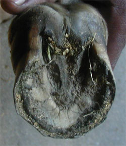 |
| Fig. 3. Medial heel displaced proximally in a foal with a rotational deformity. |
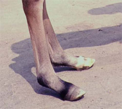 |
| Fig. 4A. Flexor flaccidly in the hind limbs of a foal showing digital hyperextension. |
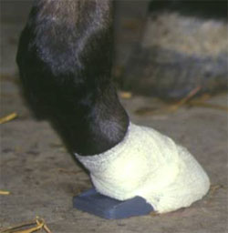 |
| Fig. 4B. Foal with plastic palmar/plantar extension shoe taped to foot. |
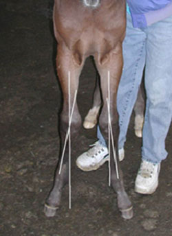 |
| Fig. 5. Carpal valgus in a foal. Note limbs deviated lateral to the midline. |
 |
| Fig. 6. Urethane extension placed on the medial side of the foot for carpal valgus. |
Rotational deformities are very common in foals (Fig. 3). For example, the narrow chest together with the relatively long limbs cause many foals to adopt a base wide stance in front which is often accompanied by outward rotation of the entire limb. This stance, which can be considered normal in foals, confers a higher degree of stability and is gradually modified as the transverse diameters of the upper body increase with growth. As the foal moves, it is quite noticeable that the outside of the hoof wall of the foot contacts the ground first as a result of the flight pattern caused by the rotated position of the limb. These foals should be trimmed level and not have their feet lowered on the outside wall. A base-wide stance in a 3 to 4 month-old foal results naturally in asymmetric hoof capsules in the frontal plane with the medial side of the hoof capsule slightly lower than the lateral aspect. If this stance is not recognized as physiological for the age and an attempt is made to "correct" it by lowering the lateral wall, there is a risk of creating an angular limb deformity where none existed previously. In cases where the medial heel bulb has been displaced proximally, the medial hoof wall is lowered slightly and if severe, composites can be used to address the hoof capsule distortion (Fig. 4). Therapeutic trimming does not offer favorable results in the malpositioned limb, as this deformity is corrected through growth. As the musculature of the chest increases, the elbows are pushed outward, rotating the limbs inward.
Flexor Tendon Flaccidity, Angular Limb and Flexural Deformities in Foals
Flexor Tendon Flaccidity
Flexor tendon flaccidity or tendon laxity is a relatively common limb deformity seen in newborn foals usually involving the hind limbs although all four limbs can be involved. Weak flexor tendons is thought to be the cause which results in digital hyperextension where weight-bearing is placed on the palmar/plantar aspect of the proximal phalanges and the toe of the hoof is raised off the ground (Fig. 5). The condition often tends to self-correct within days after birth as the foal gains strength and is allowed moderate exercise. However the tendon laxity often persists and it is not uncommon to see a fool that still has digital hyperextension at 4 weeks of age. Treatment is sequential depending on the severity of the tendon laxity and the response of the foal to treatment. Therapy begins with controlled exercise allowing the foal access to a small area with firm footing for 1 hour three times daily, the toe of the foot can be shortened and the heels can be rasped gently from the middle of the foot palmarly/plantarly to create ground surface and a palmar/plantar extension can be applied if necessary. This extension which extends approximately 3-4 centimeters beyond the bulbs of the heels immediately relieves the biomechanical instability (Fig. 6). A cuff-type extension shoea is commercially available or a small aluminum plate extension with clips. In either case, the author feels that either type of extension should be attached with adhesive tape rather than a composite if the foal is less than 3 weeks of age as this avoids excessive heat being applied to the fragile hoof capsule as the composite cures and prevents contracture of the hoof capsule at the heels. Regardless of the method of application, the extensions should be changed at 10 day intervals. Bandaging the limb is contraindicated as this will further weaken the flexor tendons.
Angular Limb Deformities
Angular limb and deformities are common limb abnormalities in foals that require early recognition and treatment.1,2,4 The pathogenesis of this problem is not clearly understood. Angular limb deformities can be classified as either congenital or acquired in the first few weeks of life. The primary lesion is an imbalance of physeal growth; for various reasons, growth proceeds faster on one side of the physis. Angular limb deformities can be further classified into two categories. Valgus (according to Webster's, it is termed valgus and varus) deformities occur when the deviation occurs lateral to the axis of the limb and varus deformities occur when the deviation is medial to the axis of the limb. The most common location of valgus angular limb deformity is the carpus while varus deformities are most often seen at the fetlock.
Carpal Valgus
It is apparent that a mild carpal valgus of 2-5 degrees offers the newborn foal a comfortable stance while nursing and eating off the ground and is considered acceptable. If the deviation exceeds 5-8 degrees then it becomes a concern and should be monitored. A few days of stall confinement on firm bedding or limited exercise in a small paddock (2-3 times a day) is a rewarding, cost-effective treatment for the early carpal valgus. Radiographs should be part of the physical examination in a foal with an angular limb deformity. Occasionally osseous abnormalities such as hypoplastic carpal bones will preclude correction of the problem without splints or a cast. Radiographs will also reveal the site and degree of deviation, and allow comparison at a later date. Conservative therapy for the management of many angular limb deformities may be successful in the newborn foal. Restricted exercise would be either strict stall confinement or brief periods of turnout in a small area with firm footing. This allows the physis to be stimulated but prevents stress and compression on the affected side of the growth plate. If the knee can be corrected by applying pressure with one hand on the inside of the knee and counter pressure with the other hand applied to the outside of the fetlock, then a splint made from polyvinylchloride (PVC) pipe fitted from the elbow to the fetlock applied for a few hours daily may be useful. A full length bandage is applied to the limb first, and then the PVC pipe is placed on the outside of the limb and secured with a bandage. This will distract the carpus laterally. The splint is often the most cost effective treatment available but must be applied with caution.
 |
| Fig. 7. Foal showing bilateral fetlock varus. |
 |
| Fig. 8. Flexural deformity. Note broken forward hoof-pastern axis and heel raised off the ground. |
 |
| Fig. 9. Composite toe extension applied to a foal with a flexural deformity. |
Acquired carpal valgus deformities can be graded from one to four according to severity (Fig. 7). Acquired carpal valgus deformities can occur anywhere from a few days onward. Mild to moderate valgus will generally respond to restricted exercise and the use of a composite extension applied to the medial side of the foot while the more severe cases require surgical intervention combined with farriery. The extension on the medial side and toward the heels appears to redirect the forces on the physis on the overloaded side of the limb by moving the plane of weight bearing toward the midline3 (Fig. 8). The extension also appears promotes centerline breakover. The extension is made from a polymethylmethacrylateb mixed with fiberglass strands applied directly to the foot and shaped to the desired width. It is trimmed like normal hoof as the foot grows distally or additional applications can be applied if necessary. In severe cases of carpal valgus where surgery is necessary, a medial extension is combined with the surgery. In many cases the surgical procedure may be performed too early before conservative therapy is allowed to correct the problem. It appears that valgus angular limb deformities involving the carpus will respond to surgery up to four months of age with full correction.
Fetlock Varus
Varus deformities involving the fetlock are common in either the front or hind limb of new born foals (Fig. 9). This deformity can be congenital or acquired within the first few weeks of life. A varus fetlock deformity requires early detection and treatment as functional closure of the distal physis of the third metatarsal/metacarpal bone is approximately twelve weeks of age. Foals with fetlock varus should have their exercise restricted and will generally respond to an extension applied to the lateral side of foot. The window of opportunity for treatment is small and the extension should be applied at 2-3 weeks of age. Again, the author is reluctant to apply a composite to a foal's foot before 2-3 weeks of age. In severe cases, surgical intervention will be necessary combined with an extension. If the foal is presented for treatment after thirty days of age, treatment becomes difficult and less effective. With both valgus and varus a deformity, the earlier treatment is instituted, the better chance of correction.
Flexural Deformities
Flexural deformities have been traditionally referred to as "contracted tendons." The primary defect is a shortening of the musculotendonous unit rather than a shortening of just the tendon portion, making "flexural deformity" the preferred term.1,4 This shortening produces a unit of functional length less than necessary for normal limb alignment of the digit resulting in fixed flexion of the various joints of the distal limb especially the distal interphalangeal joint.
Flexure deformities present at birth are thought to result from intrauterine positioning, genetics, nutritional management of the mare during gestation and the influenza virus but no causes have been proven. The congenital flexural deformity involves a combination of joints in the distal limb such that the foal assumes a "ballerina" stance with weight borne on the toes. These usually resolve in the first few days of life with repeated intervals of brief exercise in a small paddock, physical therapy and full limb bandages to relax the muscles in the forearm. If limited improvement is noted by 3 days post foaling, 3-5 gms of oxytetracycline is administered intravenously. It is repeated a second time after skipping a day if necessary.5 Toe extensions made from wood or aluminum taped on the feet may be useful. Acquired flexural deformities occur during the first four months of life and generally involve the distal interphalangeal joint (Figure 9). Acquired flexural deformities are thought to be associated with poor nutritional management in foals (increased protein, unbalanced minerals, etc.). It is this author's belief that this syndrome is not part of the developmental orthopedic disease (DOD) but rather an inherited condition that can be initiated with some form of pain response. Any discomfort in the foot or lower limb will initiate the flexor withdrawal reflex which causes flexor muscle contraction and altered position of a joint. The author has seen this scenario on numerous occasions where the feet were trimmed too short with excess sole removed causing toe bruising. The first clinical sign one may see during routine trimming is abnormal wear of the hoof wall at the toe. A closer look may reveal heat in the feet, increased digital pulse, pain on hoof testers, a prominent coronary band and an upright hoof/pastern angle. This is the time for conservative treatment: restricted exercise to decrease continued trauma, the judicious use of anti-inflammatory drugs to relieve pain, and the administration of oxytetracycline which will cause muscle relaxation, leading to normal foot-pastern alignment. At the same time, the heels are lowered gently with a rasp and a composite toe extension can be applied to the dorsal hoof wall and the fiberglass can be continued over the solar surface of the foot to protect that area from further bruising3 (Figure 3). This toe extension forms part of the foot and places continuous tension on the musculotendonous unit. If this condition is allowed to persist, it will result in irreversible changes in the foot and joint capsule requiring surgical intervention.
If the acquired flexural deformity is severe showing a marked broken forward hoof-pastern axis and the heels of the foot are raised of the ground unable to bear weight, the foal should be radiographed. The foal's forefeet are placed on two wooden blocks of equal height and the foot is radiographed with the foal as relaxed as possible but not sedated. If the radiograph reveals a marked flexural deformity involving the distal interphalangeal joint signifying a shortening in the musculotendonous unit, surgical intervention in the form of an inferior check ligament desmotomy should be performed. This is combined with lowering the heels of the foot from the widest part of the foot palmarly and applying a composite toe extension.
Discussion
Foot care in the foal requires a good working relationship between a veterinarian and a farrier. A regular trimming program for foals is essential and cost-effective when conducted as an examination and maintenance exercise. Any changes in limb angulations can be noted during these evaluations and treatment started immediately. The importance of detailed records cannot be over emphasized. Foals' feet should be trimmed appropriately paying strict to encouraging hoof mass with adequate sole to build a strong healthy foot and protect the developing structures enclosed within the hoof. Hoof care in the first few months of life is serious business and should never be taken lightly. Discussion and management with regard to feet and limbs during this period will dictate the success of the foal as a sales yearling or mature athlete. A sound foot care program is time-consuming, whereas assembly-line trimming is easy, but the former is much more beneficial.
References and Footnotes