Management of White Line DiseaseReprinted with permission from the American Association of Equine Practitioners. |
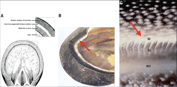 |
| Fig. 1. Illustrations of the structure of the hoof wall. (A) Hoof wall as viewed from the solar surface of the hoof. (B) Cut-away view of hoof wall showing the non-pigmented section of SM (arrow). (C) Histology slide again shows the non-pigmented section of the SM (arrow). SM, stratum medium; SI, stratum internum; SL, stratum lamallatum; WZ, white zone; S, sole. (Courtesy of Dr. Chris Pollitt). |
3. Etiology
WLD can affect a horse of any age, sex, or breed. One or multiple hoofs may be involved, and the affected hooves can be barefoot or shod. One or multiple horses on the same farm may be affected. The problem occurs worldwide. Multiple causes of WLD have been proposed, but none have been proven.
Moisture may play a role. WLD is seen more often in wet, humid areas, but there are also cases seen in hot, arid conditions. On one hand, excessive moisture softens the foot and allows for easier entry of dirt and debris into an existing separation, which can lead to a secondary infection. Continual bathing of showhorses, especially during the warmer months, may contribute to the incidence of WLD in this population of horses. On the other hand, excessively dry hooves may form cracks or separations in the hoof wall, allowing pathogens to invade.
Poor hygiene as a cause is questionable, because WLD often occurs in well-managed stables.
Infectious organisms, bacteria, fungi, or a combination of the two, have been continually incriminated as a cause. What is not known is whether these organisms are primary invaders or secondary opportunists. Given the nature of the pathogens usually isolated (mixed flora of bacteria, Pseudoallsheria and Scopulariopsis fungi), they are probably secondary opportunists, which invade and further damage an existing hoof wall separation.4 The fact that WLD can be resolved with debridement alone further detracts from this as a primary cause.1
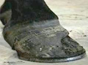 |
| Fig. 2. Long toe under-run heel as a contributing factor to WLD. Note the short shoe. |
Mechanical stress placed on the hoof wall that leads to a separation seems to be the logical cause. These stresses would include excessive toe length, poor hoof conformation, and various hoof capsule distortions such as long toe-under run heel, clubfoot, or sheared heels (Fig. 2). Damage to the stratum medium/laminar junction causes increased stress on the remaining attachment. Weight bearing and the force of the deep digital flexor tendon will cause cycling to occur, further weakening the bond.4 As the sole/wall junction becomes further damaged, it removes all remaining exterior protection, allowing for the separation to become more extensive.
Vascular damage to the hoof associated with chronic laminitis results in a compromised laminar bond and a loss of integrity (separation) at the sole/wall junction. WLD can also been noted to be a secondary problem following extensive subsolar or submural abscesses.
4. Clinical Signs
WLD offers no threat to the soundness of an animal until damage is sufficient to allow mechanical loss of the attachment between the laminae and the inner hoof wall, resulting in displacement of the distal phalanx in a distal direction (rotation and/or sinking). Only then does the horse begin to show discomfort. Most commonly, WLD is noted as an unexpected hoof-wall separation found by the farrier during routine hoof care.
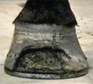 |
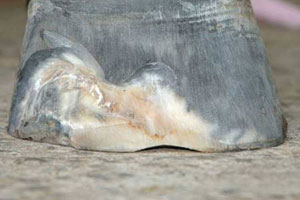
| Fig. 3. Concavity noted in the hoof wall opposite the affected area. |
In the early stages of WLD, the only noticeable change on the solar surface of the foot is a small powdery area located just dorsal to the hoof wall/sole junction. This area may remain localized, or it may progress to involve a larger area of the hoof wall. Other early warning signs of WLD may be tender soles as seen with hoof testers, occasional heat in the feet, and increasingly flat soles. As the separation becomes more extensive and extends into a quarter, a concavity ("dish") can be seen forming along one side of the hoof, and a bulge will be present on the opposite side directly above the affected area at the coronary band. There may be slow hoof-wall growth and poor consistency of hoof wall. Additionally, a hollow sound will be noted when the outer hoof wall is percussed with a hammer (Fig. 3).5 Often, the disease goes undetected until the horse begins to show discomfort.
5. Diagnosis
Lameness may or may not be observed. Hoof-tester examination does not always elicit a response. The clinical signs outlined above along with examination of the solar surface of the hoof will confirm the diagnosis. On the solar surface of the hoof, the sole/wall junction (white line) will be wider and softer and have a chalky texture. Exploration of the inner hoof wall, which lies dorsal to the white line, will generally reveal a separation filled with a white/grey powdery horn material. Further examination with a probe will reveal the depth and extent of the cavitation. There may be a black serous drainage from the separation.
If lameness is present, a thorough lameness examination should be performed to localize the suspected area. Radiographs should also be performed. With extensive hoof-wall damage, WLD accompanied by pain can mimic laminitis both clinically and radiographically.
6. Radiographs
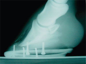 |
| Fig. 4. Radiograph showing the separation extending up the dorsal hoof wall. Note the clubfoot conformation of the foot. |
Radiology can be very informative and should be considered necessary. Good-quality radiographs consisting of a lateral view and a dorso-palmar view will show the extent of the hoof-wall separation and whether or not rotation of the third phalanx within the hoof capsule has occurred. Radiographs also allow the clinician to differentiate between WLD and laminitis (Fig. 4). Radiographically, the separation in the laminae will originate at or near the ground surface in WLD, whereas the separation will originate at the junction of the inner hoof wall and the terminal laminar papillae in laminitis. Pedal osteitis may be noted in the chronic case of WLD. Finally, radiographs will show various hoof-capsule distortions that should be addressed, and they can be used as a guide when trimming and shoeing the horse.
7. Laboratory
Laboratory findings have been unrewarding with regard to treating this disease.
Cultures may be of little value, because the samples taken from the separations are contaminated with dirt and opportunistic organisms. Aerobic cultures usually reveal a mixed bacteria flora, whereas anaerobic cultures are usually negative. Fungal cultures require a special media and time. The most common fungal species cultured are Pseudoallsheria, Scopulariopsis, and Aspergillus. A biopsy taken at the juncture between the normal and affected hoof wall shows a mixed population of microorganisms. These will generally include coccobacilli, yeast organisms, and fungal spores. Inflammation in the laminar dermis will be seen deep in the affected area.4
A technique has been described for aseptic culture of the stratum medium. This involves making a hole in the hoof wall at the proximal limit of the separated area.6 In five horses with WLD that underwent this procedure, bacterial culture was negative, but fungal culture yielded Trichoderma, Mucor, Aspergillus, or Gliocladium. These fungi are environmental inhabitants and probably are merely contaminants of an abnormal area of the hoof wall. Although this technique has proven useful for microbiological investigation of hooves with WLD, it is likely to be of limited value in a practice setting.
8. Treatment
 |
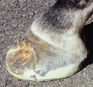
| Fig. 5. Photograph of a hoof after appropriate hoof-wall resection. There is a solid attachment of the hoof wall around the perimeter of the resection. Note that there is no hemorrhage in the resection. |
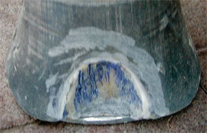 |
| Fig. 6. Tracts still present within the resection are outlined with a dye marker. |
Correction of any hoof capsule distortion that may have contributed to the hoof-wall separation is essential. Therapy of WLD is directed at protecting and unloading the damaged section of the foot with therapeutic shoeing combined with resectioning the hoof capsule that overlays the affected area.1,2,4,7,8 As a resection disrupts the continuity and weightbearing strength of the hoof wall, some type of shoe is applied to protect the hoof, to stabilize the hoof wall, and to prevent the horse from using the sole for weight bearing. If the separated area of the foot is determined to be extensive, it is important to plan the method of support and the design of the shoe before the outer hoof wall is resected. Complete hoof-wall resection (removal of outer hoof wall to expose diseased area) and debridement of all tracts and fissures in the affected area is necessary. The debridement should be continued proximally and marginally until there is a solid attachment between the hoof wall and external lamellae (Fig. 5). The veterinarian or farrier should not reach blood during debridement.
Treatment with topical medications after hoofwall resection has been described.4 Medical treatment in any form is of no value without resection of the affected hoof wall. Disinfectants/astringents such as methiolate, gention violet, or 2% iodine act as a good disinfectant but may have more benefit as a dye marker to outline the remaining tracts. The dye marker will serve as an aid in making the remaining tracts more visible at subsequent examinations and as a guide during debridement (Fig. 6). Any of the above preparations should be applied no more than twice weekly so as not to make the exposed laminae too hard. After thorough hoof-wall resection, the affected area can be left to grow out with debridement at frequent intervals. A wire brush is used daily to keep the resected area clean. Thorough exploration and debridement of any remaining tracts should take place at 2-wk intervals. When all tracts are resolved, a thorough examination is indicated at reshoeing intervals every 4 wk.
Medical treatment may not be necessary in most cases, because debridement has been proven to be sufficient. The records from 58 cases with extensive WLD treated in this practice over a 7-yr period were reviewed.1 Forty of these cases were treated with resection and continuous debridement only. The remaining cases were treated with resection, debridement, and a dye marker. In all cases, the resected portion of the foot grew out, and the hoof returned to normal. Because this disease is limited to the keratinized area of the hoof wall, this author feels that systemic medical therapy is not warranted.
The type of shoe used and the method of attachment depend on the extent of the damaged hoof wall. If the defect is small, the hoof can be shod appropriately, paying strict attention to any abnormal hoof conformation. Because the toe is involved in most cases of WLD, it is helpful to move the breakover in a palmar/plantar direction. The solar surface of the foot is trimmed from the apex of the frog palmarly/plantarly to the base of the frog, which creates two planes on the solar surface of the foot. The shoe is fitted so that breakover is placed just dorsal to the distal phalanx in an attempt to unload the dorsal hoof wall and remove the "lever arm" placed on the toe. This will also stop the "pinching" effect that often occurs at the junction of normal hoof wall and the resection.
If the resection is extensive and/or if rotation of the distal phalanx is present, a full-support bar shoe (heart bar or egg bar-heart bar combination) can be used to stabilize the foot. This type of shoe provides support to the heel area of the foot and allows some weight bearing to be transferred from the affected part of the hoof wall (toe/quarters) to the frog.
 |
| Fig. 7. A glue-on shoe allows the resection to be left open for treatment. |
An alternative method would be to use a bar shoe or open shoe combined with some type of silastic material.a The impression material can be applied to the entire solar surface of the foot as long as it was molded thicker at the heels to provide the necessary support. If no rotation is present, a good rule of thumb to follow is that if >33% of the overall surface area of the hoof is resected, use palmar/plantar support. Glueing on shoes using the ground surface of the foot may be the method of choice for shoeing the horse with WLD (Fig. 7).9 Hoof-wall separations have historically been treated by resection and acrylic repair so that nails can be placed in the affected area to attach the shoe; however, the disease process will often continue under the repair, prolonging the time required for the hoof wall to grow out. By attaching an aluminum shoe to the ground surface of the foot using a composite,b the resected area can be left open to be observed and debrided regularly. Good palmar/plantar support can also be provided with this procedure.
Acrylic repair using a polymethylmethacrylate compositeb should only be considered after all tracts are removed from the resection and the defect is solid.10 It should only be used in selected cases where the client is unable to treat the resected area and where cosmetics are a necessity. The composite may hide and/or foster infection, and it tends to weaken surrounding normal hoof wall, which can encourage reinfection. The antibiotic that is mixed with the medicated version of the composite is only effective against selected bacteria and no fungi.
9. Aftercare
A change in environment is important. The feet should be kept as dry as possible. Keeping the bedding clean and dry is helpful. Limited turnout in rain or wet weather is helpful. Turnout can be delayed in the morning until the sun can dry the dew from the pasture. A shoeing schedule should be maintained at 4-wk intervals.
Commitment from the owner with regards to a continuous treatment schedule is necessary until all signs of disease have been eliminated. The foot/feet must then be monitored monthly until the hoof wall grows out. The extent of the damage will determine the amount of time required for the treatment process; however, it is not always necessary for the horse to be out of work for this entire period of time. The amount of exercise permissible during treatment for WLD is dependent on the extent of the damage and the presence of sufficient hoof wall necessary for weight bearing.
10. Prevention
Prevention of WLD is difficult, because the exact cause is unknown. Discussing the problem with the farrier and having him/her examine each foot when the horse is shod is extremely important. Any small abnormal area involving the sole/wall junction should be explored and debrided down to solid horn. Any cavity created by debridement can be filled with medicated putty before being covered with a shoe.c Proper physiological trimming and shoeing is essential for creating a strong sole/wall junction that prevents separations and offers protection.11 Equally important is the necessity to carefully monitor horses that have previously had WLD. A year or two after WLD has been treated and resolved, it can suddenly reappear in some horses with strong hoof walls that show no sign of separation.
11. Discussion
WLD involves the inner, non-pigmented section of the stratum medium of the hoof wall, not the solewall junction (zona alba or "white line"). Thus, WLD is somewhat of a misnomer. Nevertheless, it has become the accepted term used by the majority of farriers and veterinarians. Certainly, it is a more useful term than onychomycosis, because it does not limit the primary etiology to a fungal agent.
Treating WLD has created a problem for owners, veterinarians, and farriers. Owners have been deluged with many different causes and a variety of treatments. Numerous commercially available preparations have been marketed for treating WLD, all claiming to be successful. At present, there is no convincing scientific evidence nor have there been any controlled studies performed to attest to the efficacy of these products. Veterinarians may be unaware of the magnitude of this problem, because they only see the severe cases that present for lameness evaluation and/or have apparent radiographic changes. WLD may be a subtle contributor to other causes of lameness within the foot. Farriers are very aware of this disease, because they are often confronted with nailing in compromised hoof wall or lack thereof and keeping the shoe on between resets. They continually search for medical treatments, because owners are reluctant to have sections of their horse's hoof wall removed at the farrier's recommendation. Research, owner education, and continued farrier awareness seems to be the direction of the future.