Strategies for Shoeing the Horse With Palmar Foot PainReprinted with permission from the American Association of Equine Practitioners. |
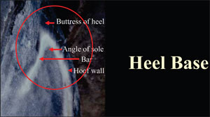 |
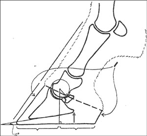
| Fig. 1. Heel base consisting of hoof wall, buttress, angle of sole, and bars. |
Damage to the Hoof Complex
The digital cushion and the heel base of the hoof capsule are two major components of the palmar foot that are susceptible to overload. The digital cushion forms the bulk of the soft tissue structures and shapes the bulbs of the heels, but when subjected to excessive stresses, it will lose structural integrity and decrease in mass. The decrease in size seems to occur through three mechanisms. First, some horses are born with genetically weak feet that fail to develop a strong palmar foot. Second, recent studies describe a period in which the cushion adapts or matures and the tissue changes from collagen bundles to fibrocartilage.6 This is a logical sequence if we consider that young horses entering training are often confined to stalls with limited or no turnout. This confinement places constant pressure on the supporting structures of the palmar foot and when coupled with limited turnout, may not provide the stimulation necessary for maturation of the cushion. During this period, the horse may receive brief periods of excessive training on immature feet, which compounds the problem. Third, if the heels of the hoof capsule are allowed to grow forward, this places a weight bearing function on the soft tissue structures of the foot, resulting in abnormal compression. Increased pressure on the digital cushion over time leads to a change in the architecture and size of the structure.
The heel base of the hoof capsule is composed of the hoof wall, heel buttress, bar, and angle of the sole (Fig. 1).2 A well-developed heel base is critical to the health of the hoof. The terms low heel or under-run heel are used interchangeably and denote the angle of the heel to be 5° or more lower than the angle of the toe, yet the structures of the heel remain intact forming a base.9 This definition may not be viable, because there are very few horses that wear shoes where the angle of the heel approximates that of the dorsal wall at the toe. Under-run heels are present when the end of the heel is located well forward of a vertical line drawn through the third metacarpal/metatarsal bone and extending to the ground. This line denotes the preferred extent of the ground surface of the heel for functional weight bearing. If the heel is allowed to grow forward, it decreases the weight-bearing surface of the foot and often precludes the heels to collapse. To formulate a rational approach to treating under-run or collapsed heels, it may be helpful to understand the process in which the heel is damaged thus showing the dilemma encountered by veterinarians and farriers when trying to improve this condition.
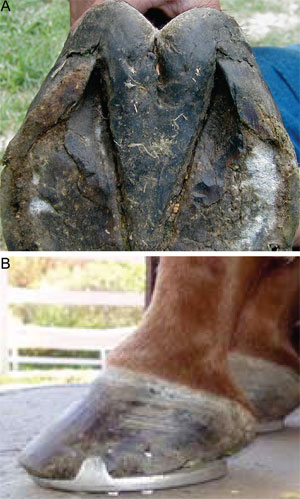 |
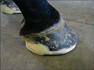
| Fig. 2. (A) Under-run heels showing damage to the heel base. (B) Lateral view of hoof capsule with under-run heels. |
The hoof wall at the heel is thin, immature horn, which makes it less rigid and therefore, more flexible compared with the thick, mature hoof wall in the toe area. This increased flexibility allows the normal physiology of the foot in the form of expansion to take place, but in turn, it makes the heels more vulnerable to damage. As this syndrome begins, the heels stop growing, and over time, the horn tubules angle forward such that the most palmar extent of the bearing surface of the hoof wall is no longer at the base of the frog but has moved forward toward the widest part of the foot. The angle of the heels become lower, and the horn tubules lose their angle and bend until they reach the point where they are parallel (horizontal) to the ground. The hoof wall at the heels becomes thinner, separates from the sole, and rolls under at the heel. The rolled under hoof wall at the heel puts pressure on the bar, which pushes it downward and laterally. This coupled with the lack of growth in the heel area makes the bar non-functional and basically nonexistent, which in turn allows the heels to become unstable, collapse, and begin to contract. As the heels grow forward, the frog and digital cushion move palmarly (Fig. 2, A and B). The heels at this point lose their mechanical strength, which prevents weight bearing. The weight-bearing function is shifted onto the soft tissue structures (frog, digital cushion, deep digital flexor tendon) in the palmar portion of the foot. Overload placed on the palmar foot leads to a loss of mass of the soft tissue structures that lead to serious consequences regarding foot function. The loss of mass allows the distal phalanx to assume a negative palmar angle, the distal interphalangeal joint is no longer able to descend and press the navicular bone into the deep digital flexor tendon during weight bearing, and the decreased surface area of the soft tissue structures places the energy of impact directly on the bone rather than dispersing it through the tissue.3
The traditional treatment for under-run heels was to use an egg bar shoe that was often accompanied by a wedge pad to raise the angle of the heels and correct the broken-back hoof-pastern axis that is usually associated with this condition. These shoes are fitted back to the bulbs of the heels with the thought being that the increased ground surface of the shoe will support the palmar section of the foot and the egg bar will support the heels, thus correcting the problem. It is questionable how one can support compromised structures (heels) that no longer have the ability to accept weight. The egg bar shoe places the bulk of the weight bearing on the soft structures of the palmar foot, which over time, will damage these structures further; the egg bar shoe further acts as a moment arm or lever on the heels, creating excessive pressure. This increased pressure will prevent hoof wall growth at the heels.
Hoof Balance
Hoof balance is a vague term used by veterinarians and farriers to describe the theoretical ideal conformation of the foot and the position of the hoof relative to the limb above.2 The term hoof balance was seldom used in early farrier texts. Hoof balance is often used during lameness evaluations to communicate aspects of therapy to the farrier rather than relying on specific biomechanical principles or therapy related to structure and function. When the term "balance the hoof" is used, multiple questions may arise, because the term has no inherent meaning. For example, "hoof balance" has been subdivided into various types such as geometric, dynamic, three dimensional, and recently, natural balance.2 Insofar as attaining overall "balance" goes, it may not be possible to satisfy all these concepts simultaneously. Geometric, dynamic, and natural balances are simply descriptive methods of hoof assessment that are used to teach the art of observation.
Geometric balance is determined by observing the horse at rest. Geometric balance implies that the dorsal, palmar, and solar view of the foot should be symmetrical. Geometric balance can also be applied using the limb axis orientation where the foot is trimmed so that the ground surface of the hoof is perpendicular to the long axis of the limb. Overall, this technique assumes that the foot and lower limb are symmetrical and that the hoof capsule reflects this symmetry. Although it is commonly used, the problem with relying on geometric balance alone is that it does not consider the mass of hoof present, the landing pattern of the foot, or the potential relationship between leg and foot conformation. Dynamic balance observes the horse in motion. This type of balance implies that a balanced foot should land symmetrically (i.e., it should land flat). By landing flat, it is hoped that the forces placed on the solar surface of the hoof wall will be uniform. The problem here is that the horse's limb conformation often precludes achieving a flat strike pattern. It may, in fact, be detrimental to the horse with abnormal upper limb conformation if it is trimmed to land in a flat strike pattern. An example of this is a horse with a rotational deformity that results in sheared heels.10
More recently, the term natural balance has been introduced. This suggests that the foot conformation of the domestic horse should be modeled after the foot in its natural state (i.e., feral horses). Obviously, the feral horse is not a domesticated athlete, and assessment of this population of horses must necessarily reflect their genetic makeup along with environmental factors that pertain to hoof wear patterns. Furthermore, it does not take into account the athletic activities of individual horses, and it is largely incompatible with traditional farriery techniques.2
Biomechanical efficiency of the hoof must be evaluated in both a dorsopalmar/plantar plane and mediolateral plane. Dorsopalmar/plantar orientation is extremely important. It ensures that the entire solar surface of the distal phalanx is loaded during weight bearing, which avoids weight bearing being concentrated on the dorsal or palmar/plantar aspects of the sole and underlying distal phalanx. Poor dorsal palmar/plantar alignment may contribute significantly to the long-toe/under-run heel conformation, the compromised heel structures, and the altered biomechanics that accompany this condition. Poor mediolateral orientation of the hoof is thought to be associated with foot problems such as sheared heels, distorted hoof walls, and hoof cracks. These problems seem to arise from disproportionate forces placed on the lateral or medial aspects of the foot during the landing phase of the stride, most often as a consequence of limb conformation.10 No one standard method of trimming/shoeing will achieve optimum foot conformation for every horse. An option to the term hoof balance would be to use a set of biomechanical principles as guidelines that could be applied to every horse. The term hoof balance could easily be replaced with hoof mechanics, which could have a universal meaning. The foot should be evaluated, trimmed, and shod in such a manner that considers the following mechanics.
Ground Surface
The usual ground surface is paramount to the effect of trimming/shoeing on the horse with palmar foot pain, and yet, it is often disregarded. During weight bearing on a hard surface, load is placed entirely on the hoof wall, but when weight bearing takes place on a deformable surface, the wall is still load bearing while the sole and frog becomes load sharing. Shifting some of the load from the hoof wall may reduce the compressive forces placed on the heel during weight bearing. A deformable surface allows the hoof to rotate about the distal interphalangeal joint, and it does not restrain movement.4 Sand surfaces will reduce loading to tendons and thus, the navicular bone and navicular ligaments.a The forward rotation of the dorsal hoof into the surface acts as a heel wedge to reduce the moment of force about the distal interphalangeal joint and thus, relieves stress on the navicular bone. Egg bar shoes have been suggested as an effective measure to reduce lameness in horses with palmar foot pain. Egg bar shoes do not reduce stress on the navicular bone on hard non-deformable surfaces but may have some effect on horses that perform on a loose, deformable surface. Egg bar shoes or any bar shoes may provide a flotation effect, because the increased ground surface of the bar does not allow the heel to readily sink into the ground.11 Rotation of the foot into a soft, deformable surface may, in fact, have the same effect as a rolled-toe, rocker-toe, or natural-balance shoe.11 If the horse is standing on a soft surface, the toe will be pressed into the soil as the deep digital flexor tendon flexes the distal interphalangeal joint. If the horse is standing or working on a hard surface, the toe has nowhere to go as the tensile forces are transferred from the distal phalanx to the dorsal hoof wall through the laminar attachment.
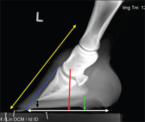 |
| Fig. 3. Lateral radiograph used to illustrate hoof-pastern axis, center of articulation, sole depth, palmar angle of distal phalanx, breakover, and placement of the shoe. |
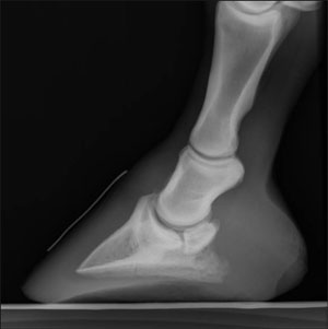 |
| Fig. 4. Lateral radiograph shows the palmar angle of the distal phalanx relative to the ground. |
Radiographs
Radiography can be used as both a diagnostic tool and an aid in assessing all structures within the foot. An unknown but commonly encountered situation is identifying the existence of foot pain in the absence of demonstrable radiographic changes. Considerable information can be obtained from the image of the overall shape of the hoof capsule, the soft tissue structures, and the position of the distal phalanx within the hoof capsule. It should be remembered that many lamenesses localized to the foot are caused by hoof capsule distortions, poor foot conformation, improper landing patterns, and soft tissue damage, which results in inappropriate biomechanical stresses being placed on the navicular apparatus and the distal interphalangeal joint. With or without radiographic lesions indicating disease, the clinician will be able to use the lateral to medial and dorsopalmar views as a precise guide to implement therapeutic trimming and shoeing. From the lateral medial view, we can readily measure the hoof-pastern axis, center of articulation, sole depth, palmar angle of the distal phalanx, breakover, and placement of the shoe (Fig. 3). When the solar plane of the distal phalanx shows a negative palmar angle, we can also access the need for heel elevation and the amount to apply (Fig. 4). The dorsopalmar view will allow the clinician to evaluate the lateral medial orientation of the distal phalanx within the hoof capsule, the position of the distal phalanx relative to the ground, and the effect of the position of the distal phalanx on the joint spaces of the digit; the clinician can then make the appropriate shoeing changes. The need to access the position of the hoof capsule relative to the long axis of the digit is often overlooked.
Concept of Therapeutic Shoeing
Therapeutic shoeing could be considered the "art and science" of affecting/influencing the structures of the foot. The scope of therapeutic shoeing can be somewhat limited if the structures of the foot are compromised, which renders the foot incapable of accepting the forces placed on the foot by a therapeutic shoe. Trimming is the "mainstay" of therapeutic shoeing, and trimming will be directly influenced by the amount of hoof mass present. Shoeing maintains what has been trimmed (protection), compliments the trimmed foot, and provides foot mechanics. The shoe is able to offer protection, increase ground surface, decrease concussion, unload areas of the foot, affect breakover, and provide heel elevation. Therapeutic shoeing that can be applied to the foot through trimming and/or shoeing can change the dorsopalmar orientation of the foot and the mediolateral orientation of the foot as well as the forces associated with breakover, the forces associated with the deep digital flexor tendon, and the amount of concussion the foot receives during impact. In the event that there is insufficient foot mass present to effect a change with trimming, mechanics can be incorporated into the shoe. Breakover, heel elevation in the form of a wedge, and a roller motion configuration are some of the mechanics that can be built into the shoe. Rails can be added to the shoe for heel elevation, and the axial placement of the rails may reduce torque on the laminar interface of the hoof capsule in a medial or lateral direction. Various forms of bar shoes offer protection, increase the ground-surface area of the foot; stabilize the vertical movement of the hoof capsule at the heels, and in some cases, offer support. Silastic materials and various anticoncussive pads can be combined with shoes to decrease concussion.
Rest
Rest or controlled exercise should, but seldom does, form part of the therapy for horses with palmar foot pain. With the advent of magnetic resonance imaging (MRI), we are able to image structures within the foot such as the deep digital flexor tendon, navicular bursa, impar ligament, and collateral ligaments. When a lesion involving one of these structures is present, enforced rest of 6-12 mo is generally required. Although the duration of the rest period associated with palmar foot pain is variable, 3-4 wk of rest or limited turnout allows the medical therapy to become effective and the soft tissue inflammation to subside; it also allows structures in the hoof complex, which have been addressed with therapeutic shoeing, to improve. A recent study showed that when evaluating the effect of shoeing, an adaptation period of 2-3 wk is necessary to achieve pain relief.11 If the lameness has resolved after a period of rest, a gradual increase in exercise is recommended before returning the horse to normal use. Although rest may be a beneficial part of treatment, owner or trainer compliance may not be forthcoming, because the desire to continue with competition may take precedent.
3. Shoeing the Horse With Palmar Heel Pain
Conventional wisdom on shoeing the horse with palmar foot pain is that each case should be regarded as an individual. It is rare to find a case of palmar foot pain with confirmed pathology in the navicular region that is not accompanied with a damaged hoof complex. Central to our current knowledge of therapeutic shoeing is the interaction of the structures of the hoof, the manner in which the foot loads, and the surface on which the horse is asked to perform. Our approach to therapeutic shoeing should address three parameters: the visual structures of the hoof complex, the function of the distal interphalangeal joint, and the areas of pathology confirmed through diagnostics and imaging. The ability to improve structures of the foot will be dependent on the structure(s) involved, the amount of damage to the structure, the duration or chronicity, its dependence on adjacent tissue, and its ability to replenish. Damage to various sections of the hoof is generally accompanied by some form of hoof-capsule distortion resulting from inappropriate loading during weight bearing.
Dorsal-palmar and medial-lateral orientations are dictated by form and placement of the distal phalanx within the hoof complex. Decrease in the angle between the palmar margin of the distal phalanx and the ground surface results in greater loading of the navicular bone, whereas differences between the toe angle/heel angles has not been correlated to increase in loading of the navicular apparatus.
 |
| Fig. 5. Schematic illustration showing the center of articulation. (Courtesy of Dr. C. Colles.) |
 |
| Fig. 6. Solar view of the foot with a line drawn across the widest part of the foot and corresponding lines drawn to the toe and the heel. |
 |
| Fig. 7. Aluminum shoe with breakover moved palmarly/plantarly. |
 |
| Fig. 8. So-called "Onion" shoe used to protect and possibly load and stimulate the bars. |
Method
To implement trimming and shoeing, guidelines are established using careful evaluation of the foot along with good quality radiographs. Trimming and shoeing techniques are applied to the foot using the biomechanical principles that address the center of articulation, the hoof pastern axis, the trimming of the heels to the base of the frog, and breakover. Trimming begins with a line drawn across the widest part of the foot that corresponds to a vertical line dropped from the center of the distal end of the second phalanx (Fig. 5).12 This line drawn across the widest part of the foot will approximate the center of articulation. Other than excessive hoof wall, no horn material is removed from the solar surface of the foot. The heels are trimmed with a rasp to the base of the frog, when possible, with the intent being to create a solid heel base. If insufficient hoof wall is present for the end of the heel to reach the base of the frog; this distance can be lengthened with the shoe. The medial or lateral wall can be lowered cautiously relative to the other when changes to the lateral-medial alignment of the foot are necessary. Excess length of the hoof wall at the toe of the hoof is determined at the sole/wall junction, and it is removed, being careful to leave the adjacent sole for protection. A line drawn from the existing line across the widest part of the foot to the base of the frog should be equal to another line drawn from the widest part of the foot to the toe (Fig. 6). This places the center of articulation in the middle of the foot or in the middle of the shoe when shod. The foot is shaped by removing flares from the outer surface of the hoof capsule to concentrate weight bearing on the hoof wall. This constitutes a basic fundamental trim to which a shoe can be applied to compliment the trim, protect what has been trimmed, and change the biomechanics of the foot further when necessary. The shoe should be as light as possible with a wide web width (0.63-0.75 in) attached with as few nails as possible of the smallest size.
Biomechanics
Biomechanics is the study of the mechanics of a biological structure.4 A working knowledge of the biomechanics of the foot as we perceive them is essential for the clinician to implement changes through farriery. Hoof mechanics are used to return the biomechanics of the foot to normal and to change the stresses exerted on structures of the foot. Various hoof mechanics are applied to the foot through the combination of trimming, shoes, composites, and urethane products. Elevation of the heels induces flexion of the distal interphalangeal joint, decreases tension in the deep digital flexor tendon, reduces pressure applied to the navicular bone, reduces stress on the hoof capsule, and decreases deformation of the hoof capsule.13-15 Heel elevation is also used to change the position of the distal phalanx within the hoof capsule (i.e., when the angle of the palmar margin of the distal phalanx is negative relative to the ground). It is the author's feeling that when possible, heel elevation should be incorporated into the shoe and placed under those structures of the foot designed to bear weight. Wedge pads or wedge inserts placed between the shoe and the foot are used to elevate the heels but often at the expense of overloading the supporting structures of the foot, which can lead to further deterioration of the hoof capsule at the heels. Wedge pads or wedge inserts should only be used if there is an adequate heel base present along with a substantial frog. Moving the breakover in a palmar/plantar direction has been shown to decrease the moment arm exerted on the distal interphalangeal joint as the foot leaves the ground, but it has not been shown to speed up the timing of breakover (Fig. 7).16 Heel elevation seems to be complimented by moving the breakover back by reducing any delay in breakover that exacerbates stress in the distal interphalangeal joint.17 Many commercial shoes are available that have heel elevation and enhanced breakover included in their design.b Various types of bar shoes are useful in the treatment of palmar foot pain. They offer protection, an increase in ground-surface area, the ability to unload areas of discomfort, stabilization of the heels in a vertical direction, and support. Support is a widely used term that is seldom defined and often ambiguous. Support as defined means to hold up a structure or to keep it from collapsing. Support as a concept applied to structures within the foot, such as collapsed heels caused by excessive compressive strain, has little overall applicability in farriery. However, supporting the foot with heel pain may be accomplished by preventing the palmar aspect of the hoof from descending into the ground surface. This can be accomplished by increasing the ground surface area around the periphery of the foot or at the heels. The author prefers a straight bar shoe over an egg bar shoe. This shoe can be fitted in close approximation to the hoof capsule, and the forces can be more concentrated under the foot, yet still accomplish the flotation effect. With the egg bar shoe, the branches of the shoe will invariably extend beyond the heels of the foot, which creates a moment arm or lever that places excess pressure on the heels of the hoof capsule.
 |
| Fig. 9. Glue-on OT shoe with breakover and heel elevation designed into the shoe. |
Protection
Protection Protection to weak, damaged, or overloaded areas of the heel can be provided by shoes or shoes combined with various silastic or urethane products. Shoes can be forged or metal plates can be attached to shoes that cover structures susceptible to damage, such as the heel base of the hoof capsule, or structures overlying the frog. Collapsed or weak heels often represent a source of pain and can be effectively protected by modifying the heels of a shoe or by attaching a small plate to the shoe that is shaped to fit over the heel base (Fig. 8). Impression materialc can be placed under the wide expanse of the shoe at the heels; it may decrease the compression placed on the heels during weight bearing, and at the same time, it may load the bars with the thought being to improve the function of the ungual cartilage. Horses with pain localized to the palmar foot often benefit from liquid polyurethaned being placed in the solar surface of the foot. This material forms a soft, deformable substance under the solar surface of the foot that increases the surface area of the foot, adds protection, decreases compression, and enhances the descent of the distal interphalangeal joint during weight bearing.
Composites
The use of acrylic composites,e not only to attach horseshoes but also to raise, reconstruct, or extend the ground surface of a weak or collapsed heel, is extensive in therapeutic shoeing.18 The composite used to glue on the shoe, when placed between the foot and the shoe at the heels, can have the following effects: elevates the heels when necessary, adds mass to the heels so that they can be loaded, extends the ground surface area of the hoof wall at the heel, and creates an interface between the shoe and the foot that will eliminate the movement of the heels against the shoe during expansion, which decreases wear of the heel structures. Glue-on shoes can be useful with horses that have palmar foot pain with decreased hoof mass because it allows the hoof mechanics to be incorporated into the shoe, and then, the shoe is placed on the foot in the proper position without the need for nails (Fig. 9).
4. Discussion
Treating hoof disease is the only area of veterinary medicine where another profession needs to be involved. A skilled, knowledgeable farrier plays a significant role in treating palmar foot pain. A working knowledge of the form and function of the equine foot is essential for equine practitioners and farriers to formulate a shoeing plan and implement the appropriate hoof mechanics. The anatomical and medical knowledge of the veterinarian, when combined with the mechanical and technical skills of the farrier, provides a comprehensive team that can only enhance the management of palmar foot pain.