HOW TO MANAGE SHEARED HEELSStephen E. O’Grady, BVSc, MRCVS Northern Virginia Equine, P.O. Box 746 , Marshall , VA 20116 Reprinted with permission from the American Association of Equine Practitioners. Original printed in the 2005 AAEP Convention proceedings
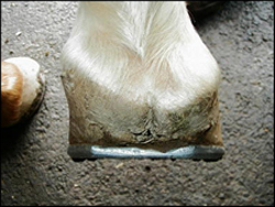
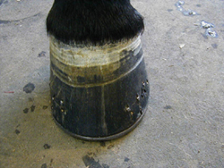
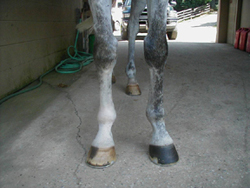
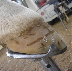
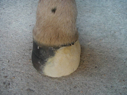
Introduction Sheared heels and its association with lameness were described in the veterinary literature thirty years ago 1. Sheared heels can be defined as a hoof capsule distortion resulting from displacement of one heel bulb proximally relative to the adjacent heel bulb (Figure 1) 2 This disparity between the lateral and medial heel bulb is generally .5 centimeters or more. When the weight of the horse is not distributed uniformly over the entire hoof during the landing phase of the stride, one area of the foot, usually a heel or heel and accompanying quarter, receives a disproportionate amount of the total force associated with impact. This resultant force leads to a remodeling of the affected heel bulb. The degree of distortion in the affected heel is dependent upon the amount of concussion sustained by the individual foot. Sheared heels can occur in the hind feet as well as the forefeet (Figure 2).
Causes that have been described for sheared heels are:
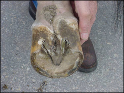
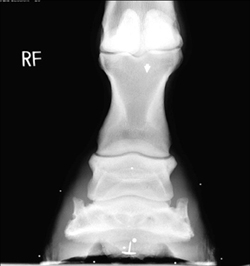
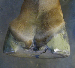
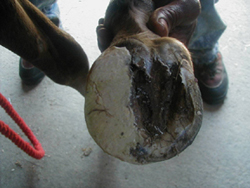
Although a number of sound horses have distorted hoof capsules, lameness is often attributed to this condition. This continual disproportionate loading and increased compressive stresses on one heel/quarter predisposes the foot to hoof capsule distortion, subsolar bruising, corns, quarter and heel cracks, fracture of the bar, deep fissures within the base of the frog and thrush in narrow frogs 4. In fact, seldom is one of the above conditions present when not accompanied by a sheared, contracted or under-run heel. This paper will review the etiology, purported causes and the current treatment strategies for sheared heels. STRUCTURAL CHANGES TO THE FOOT The equine hoof capsule is a viscoelastic structure that has the unique ability to deform when weight is accepted uniformly. However, if the energy of impact is continually placed on one side of the foot along with abnormal loading on the other side, over time, structural changes will become apparent. The increased concussive forces on one side of the foot cause the hoof wall to assume a steeper angle, that is, the wall becomes straighter. Along with the increased hoof angle, other changes such as contracture of the heel subjected to the greater forces will soon follow. This decreases the ground surface of the foot, resulting in a lack of expansion on that side and makes the solar surface in the palmar/plantar section of the foot asymmetrical. This asymmetry is easily seen if one considers that the frog normally bisects the hoof. Over time, the hoof wall begins to “roll under” on the affected side, which further decreases ground surface under that area of the foot. The lamina above the hoof wall on the affected side are subjected to abnormal vertical forces that result in hemorrhage, stretching or tearing. The side of the foot that first impacts the ground develops a flare due to bending of the hoof tubules. With the abnormal strike pattern associated with sheared heels, the distal interphalangeal, pastern and fetlock joints are likely to be loaded unevenly. In addition to the uneven loading on the joints, a rotational torque is created around the point where the foot first contacts the ground, transferring this torque up the limb and placing undue stress on the joints above. This type of landing pattern also places abnormal concussive forces on the navicular bone and its associated structures. The unequal distribution of vertical forces on a given side of the foot over time results in biological remodeling rather than the heel being “pushed” proximally, i.e. the heel is “growing out of shape rather than being pushed out of shape” 3. Remodeling is the response to soft tissue deformation over time. Increased hoof wall growth could result from stimulation of the soft tissue from continuous impact, receptors in the soft tissue and the increased blood supply in the heel region. Displacement of the soft tissue due to mechanical stress would also play a role. To substantiate this statement, a horizontal dorsopalmar (DP) radiograph on fifty horses with a foot with one heel bulb that was displaced proximally .5 centimeters or greater was reviewed by the author. In all cases, it was found that the solar surface of the third phalanx was horizontal (parallel) with the ground indicating the disparity in heel height was not originating from the hoof wall located beneath the third phalanx in the heel (Figure 3).
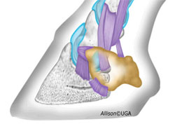
In addition, the third phalanx occupies a small portion of the hoof capsule in the heel area with the majority of space being occupied by soft tissue (Figure 4). This may account for the proliferation of soft tissue and the additional hoof wall growth occurring above the ground surface of the foot.
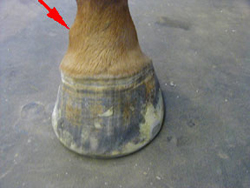
ETIOLOGY In order to formulate a rational approach to management, it is necessary to discuss the etiology of sheared heels. Sheared heels can be acquired or conformational. Improper trimming and shoeing have been considered to be common cause of sheared heels. During routine hoof care by the farrier, unequal hoof wall removal from one heel can lead to an abnormal mediolateral orientation of the hoof. It’s reasonable to assume that trimming the heels unevenly during routine hoof care would cause sheared heels; however, the viscoelastic nature of the hoof capsule could negate this effect in a well conformed limb. The author performed a small field study using a group of 25 horses (10 broodmares and 15 young horses in training) on a large breeding farm to test this hypothesis 4. The young horses were in work and half were shod and half were barefoot. To enter the study, all horses had to land flat on both forefeet before trimming their feet. One side of the foot was lowered relative to the opposing heel every four weeks for three months and in no instance was a sheared heel created. With the increased awareness of foot problems by the horse-owning public and with the continued improvement in the quality of horseshoeing, improper trimming of the hoof may not be the most common cause of this condition today. Sheared heels may arise from attempts to alter conformation by trimming or shoeing. This is done in an attempt to alter gait for performance reasons or to compensate for faulty conformation or to deceive a would-be purchaser of a horse. In trying to correct toe-out conformation especially in young horses, the lateral quarter and heel are often lowered and the medial heel is left high instead of trimming the foot level (Figure 5).
The result may show cosmetic improvement when the horse stands, but when moving; the arc of flight may be changed leading to an altered landing phase. When trimmed in this manner, the ground surface of the inside of the foot is decreased in length relative to the ground surface of the outside of the foot. In many instances, when a shoe is then applied, the branches of the shoe will be unequal in length, thus decreasing the support and increasing the force of impact on the shorter side. Conformational faults in the upper limb that change the horse’s flight phase of the stride can result in unequal loading of the foot as it strikes the ground. In this instance, the altered flight pattern causes the horse to impact the ground with one side of the foot prior to loading the foot toward the opposite quarter/heel of the foot. This excessive load displaces the heel bulb proximally, creating the unequal heel height. In the conformationally predisposed horse, the carpus is generally rotated laterally. When viewed from the front, although the entire limb faces outward, or in some instances, medially, the limb from the carpus to the ground surface of the foot forms a straight line indicating a rotational deviation of the limb (Figure 6).
With the carpus facing outward, breakover takes place in this direction, changing the flight of the foot during the stride so that the foot is unable to land evenly on both heels. As the horse approaches the landing phase of the stride, this flight pattern forces the foot to impact the ground on one side of the foot and then sustain excessive loading on the opposite side. Using a slow motion video camera, one can actually distinguish the point where the foot impacts the ground on one side and where the hoof accepts the load across the surface of the foot toward the other side. Furthermore, there appears to be a correlation between an offset third phalanx and sheared heels (Figure 7a, 7b).
Most commonly the third phalanx is offset laterally within the hoof capsule rather than directly under the first and second phalanges with the ensuing concussive forces placed medially causing the medial heel to displace proximally. Foals may develop a sheared heel at an early age due to a rotational deformity of the forelimbs. Again, this rotational deformity changes the flight portion of the stride causing the foal to land on one side of the foot instead of landing flat. Lastly, such hoof capsular distortion may be the result of chronic lameness and an associated change in gait. Standardbred racehorses with long term degenerative joint disease (DJD) involving the distal tarsus (bone spavin) will often demonstrate hoof capsule changes such as remodeling of the medial heel, manually detractable heels and a flared lateral toe 5 DIAGNOSIS The evaluation of sheared heels begins with visual assessment of hoof and limb conformation with the horse standing on a hard level surface. The gross changes noted in the foot are proportional to the amount of impact sustained, the extent of structural damage and the duration of the condition. When sheared heels are present, the heel bulb on the affected side is displaced proximally when viewed from behind the horse. When viewed from the front, the hoof wall on the affected side is straighter and, in chronic cases, will begin to roll under the horse. There is a marked flare of the hoof wall at the diagonal toe/quarter present on the side opposite the affected heel. When viewed from the side, the coronary band is displaced proximally above the damaged heel instead of having a gradual slope from the toe to the heel. The solar surface of the foot reflects changes elsewhere in the hoof capsule. The foot will be less symmetrical; the sole will appear wider on the side with the flare and narrower on the side with the affected heel. The frog is usually narrow and there is often a deep fissure located at the base of the frog that may extend to the hairline and in severe cases, when a heel is held in each hand, the heels can often be manually moved in opposing directions. The horse may show discomfort when this manipulation is performed. It is important to view the horse in motion on a hard level surface from the front and rear. This should be done at a walk and a trot. When viewed from behind, this should determine which part of the foot is impacting the ground and which portion of the foot is receiving the abnormal load. The direction of breakover should be noted when viewed from the front. Breakover will generally take place to the side of the foot with the affected heel. If lameness is present and the distorted hoof is thought to be the cause, the pain should be localized to the suspected area using hoof testers, diagnostic local anesthesia and radiology. It should be determined whether the lameness is due to or related to the sheared heels, or if another problem is present. MANAGEMENT OF SHEARED HEELSAdult horses Selective trimming coupled with therapeutic shoeing attempts to decrease the impact on the distorted heel by altering the strike pattern. Hoof trimming should be directed toward improving the landing pattern of the hoof rather than trimming the hoof perpendicular to the long axis of the limb 6,7. The latter trimming pattern does not take into consideration any conformational faults. Confusion exists as to which side of the hoof should be lowered. When a DP radiograph of a foot with a sheared heel is examined, there may be a narrowing of the proximal and distal interphalangeal joints on the side with the elevated heel bulb and the middle phalanx appears to slide toward the lower side. It will be further noted on the radiograph that the length of the hoof wall on the side of the sheared heel is longer. These two findings are evidence that the hoof wall length should be decreased on the affected side. Clinically, although not fully understood, trimming the heel on the affected side lower allows the horse to assume a more even (flatter) strike pattern. The author prefers to trim the foot in stages each of which is followed by watching the horse walk on a hard flat surface. Before trimming, the foot is lifted off the ground, the metacarpus is held horizontally and the limb is allowed to hang in its natural position under the horse. The examiner’s head is positioned over the foot so as to be able to sight along the limb and down across the solar surface of the foot, evaluating the mediolateral orientation relative to the ground. The foot is then lightly trimmed until it is as level as possible. The length of the affected heel may then approximate the length of the opposite heel and the end of the heel will be closer to the base of the frog. No more hoof wall is removed than is required to level the solar surface of the foot. The horse is then walked and the strike pattern noted. Continue to trim the affected side until the landing pattern is improved. Both heels generally will not be the same height when finished. Any flares on the opposite side of the foot are removed by rasping the outer hoof wall. Wide web steel straight bar shoe is fitted as symmetrically as possible underneath the long axis of the limb using the apex of the frog as a central marker 8. The bar shoe effectively increases the surface area of the foot, provides more expansion (ground surface) on the side with the straighter wall, and decreases the vertical movement of the heel bulbs. Wide web aluminum bar shoes can be used if the athletic endeavor of the horse dictates their use. Before applying the shoe, any remaining hoof wall under the affected heel that can be safely removed is trimmed away. This creates a space between the heel and the shoe that allows the displaced heel to drop down and settle into a more normal configuration (Figure 8).
It may require several resets using the method described above to achieve (when possible) symmetrical heel positions. Although selective trimming accompanied by some form of rigid support shoe is the treatment of choice, this method may not always change the conformation of the foot sufficiently if the heel is severely distorted. In this case, the author has successfully used another procedure prior to shoeing that provides a gross anatomical change in the affected portion of the heel prior to shoeing 9. The procedure begins by removing the shoe. Any excess sole is removed and the feet are soaked in hot water kept at a constant temperature for twenty minutes. A frog support pad a is taped to the bottom of the foot and a heavy cotton bandage is applied to surround the entire foot including the coronary band. The horse is placed in a stall for 24 hours and the bandage is moistened with hot water periodically during that time. Alternatively, following the initial soak, the foot could be wrapped in a self-contained moist poultice b for 24 hours. Keeping the foot moist renders the hoof capsule more pliable so that movement of the hoof capsule toward a more normal physiologic shape can take place around a central focus which is the supported coffin bone. The following day, when the bandage is removed, the distorted heel will have assumed a more normal position depending on the severity of the condition at the onset. The foot (feet) are now trimmed and shod as described previously. Foals Sheared heels in foals are generally the result of a rotational deformity of the forelimbs often combined with improper trimming. These foals stand toed-out and generally have their outside hoof wall trimmed lower regardless of the cause of their limb position. If this toed-out stance is a result of outward rotation of the knees coupled with a narrow chest, trimming in this manner compounds the problem. Improving the sheared heel involves gradually trimming the foals hoof level. The correction is done gradually and the affected side is lowered a few millimeters each time the foal is trimmed generally at 4 week intervals. If the condition is severe, the medial heel will be displaced proximally and it will have begun to roll under causing a reduction in the ground surface of the hoof. In this case, an extension to the hoof is fabricated using a composite c material mixed with strands of fiberglass and is attached to the side of the affected quarter/heel. The extension will increase the width of the hoof wall (Figure 9a, 9b).
The solar surface of the foot is not raised with the extension, this serves two purposes. First, it causes the foal to break over straighter, improving the limb flight and resulting in a more uniform strike pattern. Second, the extension prevents further bending of the hoof wall, and adds ground surface to the heel. As the hoof wall grows distally, it will in many cases follow the direction of the composite so that the wall at the heel will bend less. A large number of these foals will improve as they grow because, as the chest widens, the rotational deformity improves, changing the landing pattern. DiscussionThe prognosis for this condition is good provided a skilled interested farrier is involved. It is also necessary to have a committed owner as resolution (when possible) of these cases is slow and they often require ongoing maintenance. Theoretically, the prevention and treatment of lameness caused by inappropriate mediolateral orientation is simple but in practice it is often difficult to achieve. Being aware that sheared heels can predispose the horse to multiple hoof wall problems makes prevention and treatment imperative. When lameness is associated with sheared heels, treatment becomes necessary. However sound horses with a sheared heel from whatever cause also will benefit from correction. Many times, improvement is all that can or will be achieved. References
Footnotesa Lily Pad, Advanced Equine Productions. PO Box 54 , Versailles KY 90383 . b Animalintex, ® 3M Animal Care Products Division. 3M Center Building , St. Paul , MN 55144 c Equilox, ® Equilox International, 110 2 nd Street NE , Pine Island , MN 55963 . |