How to Treat Equine CankerStephen E. O'Grady, BVSc, MRCVS, John B. Madison, VMD, Diplomate ACVS †
Reprinted with permission from the American Association of Equine Practitioners. Original printed in the 2004 AAEP Convention proceedings
Take-Home Message: The incidence of equine canker appears to be more prevalent than once thought. The treatments outlined in the literature for this disease are sparse, varied and generally ineffective. The authors outline a treatment protocol for canker, that when coupled with owner compliance, will resolve the disease. Introduction Equine canker is described as an infectious process that results in the development of a chronic hypertrophy of the horn-producing tissues.1 It generally originates in the frog; may remain focal, but has the capacity to become diffuse and invade the adjacent sole, bars and hoof wall. Canker can occur in one foot or multiple feet may be involved. The disease is commonly seen in draft breeds but can affect any breed or sex. Recently, one author (SEO) has seen severe canker in two imported Warmblood horses. The etiology of canker remains elusive but wet environmental or moist unhygienic conditions have traditionally been thought to act as a stimulus, however, canker is commonly seen in horses that are well cared for and horses who receive regular hoof care. One author (JBM) observed a seasonal incidence of canker in Florida as the majority of cases presented to his hospital were during the months of July through December. The treatments described in the literature have consisted of debridement and the application of topical medications including antibiotics, astringents, antiseptics, and caustic powders. No treatment to date has been consistently effective in treating this disease and the prognosis has always been guarded...
Canker generally originates in the frog and can be mistaken for thrush in the early stages. Thrush is limited to the lateral and medial sulci or the base of the frog if a fissure is present whereas canker invades the horn of the frog anywhere throughout its structure. There is a proliferation of tissue with canker versus a loss of tissue as with thrush. In the early stages canker may present as a focal area of granulation tissue in the frog that bleeds easily when abraded. Upon closer inspection a light brown or grey tissue will surround this focal area (Fig 1). 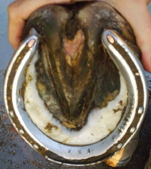
If left untreated, the disease will become diffuse and involve the frog, bars, sole and the stratum medium of the hoof wall in the palmar/plantar aspect of the foot. The infection results in abnormal keratin production or dyskeratosis, which is seen as filamentous fronds of hypertrophic horn.2 Canker is characterized by numerous small finger-like papillae of soft off-white material that resembles a cauliflower-like appearance (Fig 2). 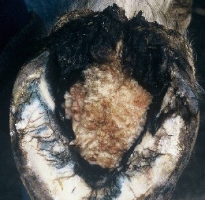
1 The condition is frequently but not always accompanied by a foul odor and is covered with a caseous white exudate that resembles cottage cheese (Fig 3). 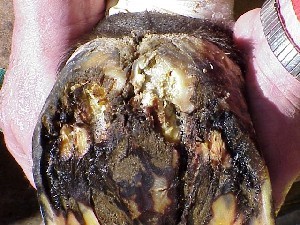
The frog is often undermined with the horny frog covering the bulk of the disease. The affected tissue will bleed easily when abraded and may be extremely painful when touched. Varying degrees of lameness will be present depending on the extent and depth of the infection. Most horses are not lame when the disease is recognized and treated early. The presence of lameness frequently indicates that the disease involves more than the superficial horny frog and warrants an aggressive approach to resolving the problem.
A presumptive diagnosis of canker is based on the gross appearance of the affected horny tissue along with a fetid odor; however a definitive diagnosis may be confirmed with a biopsy. Biopsy is most useful in recurrent cases or when the lesions do not have the characteristic appearance or they appear in unusual locations of the foot. Care must be taken to remove the superficial necrotic tissue before the biopsy is taken from the margin of the lesion. The biopsy should include both normal and abnormal tissue.3 A 6 mm biopsy punch works well. Histologically, the lesion is read as a chronic, hypertrophic, moist pododermatitis of the frog. It is characterized by a proliferative papillary hyperplasia of the epidermis with dyskeratosis, keratolysis and ballooning degeneration of the outer layers of the epidermis. A mixed population of bacterial organisms are observed in the stratum germinativum layer of the epidermis of the frog.2 Cultures per se are unrewarding as they typically produce an assortment of environmental organisms, Bacteroides sp.and Fusobacterium necrophorum. 3, 4
Canker always carries a guarded prognosis but recently these authors have been successful with the following approach. Treatment consists of thorough careful debridement of the affected tissue followed by a regimen of topical therapy applied daily and continued until the disease is resolved. To debride the affected tissue, the horse can be placed under general anesthesia or regional anesthesia can be used with the horse standing. The horse?s foot is trimmed appropriately removing all loose exfoliating sole as well as any excess toe or heel. The use of a tourniquet is essential. Firm pressure is placed across the vascular bundles over the abaxial surface of the sesamoids using either an Esmarch a bandage or simply a few tight turns of a cohesive bandage. Debridement can be performed in two ways. One author (JBM) uses electric cautery with the horse under general anesthesia while the other author (SEO) uses a sharp hoof knife and a number 12 scalpel blade followed by cryotherapy with the horse standing. All abnormal tissue is removed down to normal corium. A clear demarcation will be seen between normal and abnormal tissue. It is important not to remove excessive corium as this will retard cornification following surgery and may decrease the quality and depth of new sole being produced. It may be helpful to remove 1-2cm of normal tissue around the wound margins to ensure all abnormal tissue is removed. 5 If the decision has been made to place the horse under general anesthesia, use of a typical cautery handle in the cut mode allows accurate excision of hoof tissue including frog and normal horny sole. The handle used in this way will rapidly cut through sole and frog leaving only a dry eschar behind. The cautery tip may be bent as needed to undermine the base of the mass. Debridement can be carried out in the same manner using a sharp hoof knife and is followed by cryotherapy to freeze the area that has been debrided. Liquid nitrogen has always been used for this purpose but another practical method is to freeze the debrided area with a coolant spray b that is available for electrical circuits. The area of the foot that has been debrided will be soft and pliable. Freeze this affected area until the tissue becomes hard (known as hard freeze), allow the area to thaw and then repeat the freeze once more. Gauze 4 x 4 sponges soaked in a solution of 10% benzoyl peroxide in acetone c and sprinkled with a fine powder made by crushing metronidazole tablets with a mortar and pestle or a pill grinder are then packed in the defect. In large defects, to help insure contact of the medication in the depths of the wound and to minimize the production of exuberant granulation tissue, a putty elastomer material is used to form an insert to fit in the bottom of the foot (Fig 4). 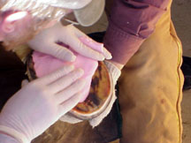
The impression material should not extend below the bearing surface of the hoof wall as this will create excessive pressure and make the horse sore. The foot is then bandaged with a dry bandage. The affected area is cleaned daily with surgical scrub, rinsed with saline, dried with a paper towel and the topical medication reapplied. It is crucial to keep the animal in a dry environment. A shoe with a treatment plate can also be used but it is sometimes hard to keep the foot as dry as necessary with this method. The authors prefer the use of bandages. Small reoccurrences may be managed with the horse standing and local anesthesia using either laser photoablation or cryotherapy. The use of systemic antibiotics such as chloramphenicol or oxytetracycline have been advocated but these authors question the use as the cases treated have resolved with local treatment only.4 A commitment is necessary from the owners, as aftercare will take several weeks to months depending on the stage of the disease until the affected tissue is cornified (Fig 5). 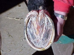
Results The records of 56 cases of canker that were treated with the above protocol from 1998 - 2004 were reviewed. The affected limb(s) were recorded in 54 cases. There were 21 with forelimb involvement, 29 with hindlimb involvement and one horse with one forelimb and one hindlimb limb affected. Five horses were affected bilaterally. Three horses were affected in all four limbs. Only one reoccurrence was recorded and that horse was treated with laser photoablation. Two horses are still undergoing treatment.
The treatment of equine canker has always presented a dilemma for veterinarians and farriers due to the poor prognosis. The etiology of canker remains obscure; however, the disease as seen by the authors differs in some respects from the disease that was described in the old surgical texts. It does not appear to be a disease of poorly cared for horses. In fact most of the horses in the study were well cared for and received routine hoof care. While the hindlimbs seemed to be affected more frequently, forelimb involvement is common. In the majority of cases, the condition starts on the frog near the heel lateral or medial to the sulcus. From that point, it can extend anywhere in the foot and even break through the hoof capsule.
|