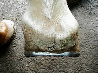
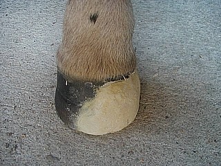
 Sheared heels can be defined as a hoof capsule distortion resulting from displacement of one heel bulb proximally relative to the adjacent heel bulb (Figure 1). When the weight of the horse is not distributed uniformly over the entire hoof during the landing phase of the stride, one focal area of the foot, usually the heel or heel and accompanying quarter, receives a disproportionate amount of the total force of impact. The resultant force leads to a structural breakdown between the heel bulbs. The degree of deformity in the affected heel (heels) varies depending on the amount of impact sustained by the individual foot. Sheared heels are diagnosed by a disparity between the medial and lateral heel lengths of 0.5 cm or more. Lameness has been attributed to this condition but a number of sound horses also have distorted hoof capsules. 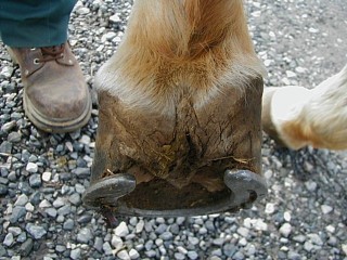 However, of equal importance, this continual disproportionate impact and increased compressive stresses on one heel predisposes the foot to pain, subsolar bruising, corns, quarter and heel cracks, fracture of the bar, pedal osteitis, deep fissures within the base of the frog, and thrush in narrow frogs. In fact, seldom is one of the above conditions present when not accompanied by a sheared, contracted or under-run heel. The management of sheared heels is through appropriate trimming and shoeing aimed at restoration of proper heel alignment. Sheared heels can occur in the hind feet as well as the forefeet (Figure 2). STRUCTURAL CHANGES TO THE FOOT
The equine hoof capsule is a viscoelastic structure that has the unique ability to deform when weight is accepted uniformly. However, if the energy of impact is continually placed on one heel, over time, structural changes will become apparent. The increased focal impact on one side of the foot causes the hoof wall to assume a steeper angle, that is, the wall becomes straighter. Along with the increased hoof angle, contracture of the heel subjected to the greater forces will soon follow. This decreases the ground surface of the foot, resulting in a lack of expansion on that side and makes the solar surface of the foot asymmetrical. This asymmetry is easily seen if one considers that the frog normally bisects the hoof. Over time, the hoof wall begins to "roll under" on the affected side, which further decreases support under that area of the foot. The laminae above the hoof wall on the affected side are subjected to abnormal shearing forces that result in hemorrhage, stretching or tearing. The side of the foot that first contacts the ground develops a flare due to bending of the hoof tubules. With the abnormal strike pattern associated with sheared heels, the distal interphalangeal, pastern and fetlock joints are loaded unevenly. In addition to the uneven loading on the joints, a rotational torque is created around the point where the foot first contacts the ground. This torque is transferred up the limb and places undue stress on joints, ligaments and the suspensory apparatus. This type of landing pattern also places abnormal concussive forces on the navicular bone and its associated ligaments. ETIOLOGY
In order to formulate a rational approach to therapeutic shoeing, it is necessary to discuss the etiology of sheared heels. Sheared heels can be conformational or acquired. Conformational faults in the upper limb that change the horse's flight phase of the stride can result in unequal loading of the foot as it strikes the ground. In this instance, the altered flight pattern causes the horse to contact the ground with one side of the foot prior to impact on the opposite heel followed by full weight bearing on that side of the foot. This focal impact drives the heel proximally, creating the unequal heel height. In the conformationally predisposed horse, the carpus is generally rotated laterally but occasionally medially. When viewed from the front, although the entire limb faces outward, or in some instances, medially, the limb from the knee to the ground surface of the foot forms a straight line indicating a rotational deviation of the limb. With the knee facing outward, breakover takes place in this direction, changing the flight of the foot during the stride so that the foot is unable to land evenly on both heels. As the horse approaches the landing phase of the stride, this flight pattern forces the foot to contact the ground on one side of the foot and then sustain excessive impact on the opposite side. Using a slow motion video camera, one can actually distinguish the point where the foot contacts the ground on one side and the point where the hoof impacts the surface on the other side. Furthermore, there appears to be a correlation between an offset third phalanx and sheared heels. Most commonly the third phalanx is offset laterally within the hoof capsule rather than directly under the first and second phalanges with the ensuing concussive forces causing the medial heel to shear.
Foals may develop a sheared heel at an early age due to a rotational deformity of the forelimbs. Again, this rotational deformity changes the flight portion of the stride causing the foal to land on one side of the foot instead of landing flat.
Improper trimming and shoeing have been considered to be the most common cause of sheared heels. This scenario is different from the first cause as the landing pattern is created by human intervention. Excess hoof wall is removed from one heel leading to an abnormal mediolateral orientation of the hoof. Improper or poor trimming may be unintentional because the farrier does not understand the goals of maintaining proper mediolateral orientation or is unable to implement these goals. Because the heels are a different length, a disproportionate force is placed on the longer heel during weight bearing. This is thought to cause an abnormal shearing force between the heels and structural breakdown with the affected heel being driven upward. However, the author used a group of 20 horses (10 broodmares and 10 young riding horses) on a large breeding farm to test the latter hypothesis. All horses landed flat before trimming their feet. I repeatedly lowered one side of the foot, sometimes excessively and in no instance was I able to cause sheared heels. With the increased awareness of foot problems by the horse-owning public and with the continued improvement in the quality of horseshoeing, improper trimming of the hoof may not be the main cause of this condition today.
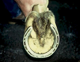 Sheared heels can also arise from attempts to alter conformation by trimming or shoeing. This is done in an attempt to improve performance or to compensate for faulty conformation or to deceive a would-be purchaser of a horse. In trying to correct toe-out conformation especially in young horses, the lateral quarter and heel are often lowered and the medial heel is left high instead of trimming the foot level. The result may show cosmetic improvement when the horse stands, but when moving; the arc of flight may be changed leading to an altered landing phase. When trimmed in this manner, the ground surface of the inside of the foot is decreased in length relative to the ground surface of the outside of the foot. In many instances, when a shoe is then applied, the branches of the shoe will be unequal in length, thus decreasing the support and increasing the force of impact on the shorter side (Figure 3). Sheared heels can also arise from attempts to alter conformation by trimming or shoeing. This is done in an attempt to improve performance or to compensate for faulty conformation or to deceive a would-be purchaser of a horse. In trying to correct toe-out conformation especially in young horses, the lateral quarter and heel are often lowered and the medial heel is left high instead of trimming the foot level. The result may show cosmetic improvement when the horse stands, but when moving; the arc of flight may be changed leading to an altered landing phase. When trimmed in this manner, the ground surface of the inside of the foot is decreased in length relative to the ground surface of the outside of the foot. In many instances, when a shoe is then applied, the branches of the shoe will be unequal in length, thus decreasing the support and increasing the force of impact on the shorter side (Figure 3).
Traction devices such as "stickers" placed on one heel will concentrate the energy of impact to that focal area of the foot. This single heel calk will cause elevation of one heel, cause the foot to tilt and change the mediolateral orientation of the foot. Continued use of this type of traction device often leads to the heel being displaced proximally. DIAGNOSIS
The evaluation of sheared heels begins with visual assessment of hoof and limb conformation with the horse standing on a hard level surface. The gross changes noted in the foot are proportional to the amount of impact sustained, the extent of structural damage and the duration of the condition. When sheared heels are present, the heel bulb on the affected side is displaced proximally when viewed from behind the horse. When viewed from the front, the hoof wall on the affected side is straighter and, in chronic cases, will begin to roll under the horse. There is a marked flare of the hoof wall present on the side opposite the affected heel. When viewed from the side, the coronary band is displaced proximally above the damaged heel instead of being parallel with the ground. The solar surface of the foot reflects changes elsewhere in the hoof capsule. The foot will be less symmetrical; the sole will appear wider on the side with the flare and narrower on the side with the underrun wall. There is often a deep fissure located at the base of the frog that may extend to the hairline and in severe cases, when a heel is held in each hand, the heels can be manually moved in opposing directions. The horse may show discomfort when this manipulation is performed.
It is important to view the horse in motion, again on a hard level surface from the front and rear. This should be done at a walk and a trot. When viewed from behind, this should determine which part of the foot is contacting the ground and which portion of the foot is receiving the impact. The direction of breakover should be noted when viewed from the front. Breakover will generally take place to the side of the foot with the affected heel.
If lameness is present and the distorted hoof is thought to be the cause, the pain should be localized to the suspected area using hoof testers, diagnostic local anesthesia and radiology. It should be determined whether the lameness is due to or related to the sheared heels, or if another problem is present. CORRECTIVE SHOEING
Adult horses
Corrective shoeing coupled with selective trimming of the hoof attempts to decrease the impact on the distorted heel by altering the strike pattern. Hoof trimming should improve the landing pattern of the hoof rather than trimming the hoof perpendicular to the long axis of the limb. The latter trimming pattern does not take into consideration any conformational faults. Confusion exists as to which side of the hoof should be lowered. When an A-P radiograph of a foot with sheared heels is examined, there is a narrowing of the proximal and distal interphalangeal joints on the side of hoof elevation and the middle phalanx slides to the lower side. It will be further noted on the radiograph that the length of the hoof wall on the side of the sheared heel is longer. These two findings are ample evidence that the hoof wall length should be decreased on the affected side. Clinically, after trimming the heel on the affected side lower, the horse will have a more even (flatter) strike pattern.
The author prefers to trim the foot in stages each of which is followed by watching the horse walk on a hard flat surface. Before trimming, the foot is lifted off the ground, the metacarpus is held horizontally and the limb is allowed to hang in its natural position under the horse. The examiner's head is positioned over the foot so as to be able to sight along the limb and down across the solar surface of the foot, evaluating the mediolateral orientation relative to the ground. The affected heel is usually high (longer) when viewed in this manner. The foot is then trimmed level which entails removing more hoof wall on the affected side. The length of the affected heel will then approximate the length of the opposite heel and the end of the heel will be closer to the base of the frog. No more hoof wall is removed than is required to level the solar surface of the foot. However, additional hoof wall may be removed on the affected side if necessary to achieve the desired strike pattern. Any flares on the opposite side of the foot are removed by rasping the outer hoof wall.
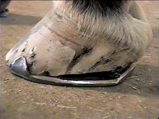 A wide web steel straight bar shoe is fitted as symmetrically as possible underneath the long axis of the limb using the apex of the frog as a central marker. The bar shoe effectively increases the surface area of the foot, provides more expansion (support) on the side with the straighter wall, and stops the vertical movement of the heel bulbs. Wide web aluminum bar shoes can be used if the athletic endeavor of the horse dictates their use. Before applying the shoe, any remaining hoof wall under the affected heel that can be safely removed is trimmed away. This creates a space between the heel and the shoe that allows the displaced heel to drop down and settle into a more normal configuration (Figure 4). It may require several resets using the method described above to achieve (if possible) symmetrical heel positions. A wide web steel straight bar shoe is fitted as symmetrically as possible underneath the long axis of the limb using the apex of the frog as a central marker. The bar shoe effectively increases the surface area of the foot, provides more expansion (support) on the side with the straighter wall, and stops the vertical movement of the heel bulbs. Wide web aluminum bar shoes can be used if the athletic endeavor of the horse dictates their use. Before applying the shoe, any remaining hoof wall under the affected heel that can be safely removed is trimmed away. This creates a space between the heel and the shoe that allows the displaced heel to drop down and settle into a more normal configuration (Figure 4). It may require several resets using the method described above to achieve (if possible) symmetrical heel positions.
Although selective trimming accompanied by some form of rigid support shoe is the treatment of choice, this method does not always change the conformation of the foot if the heel is severely distorted. This author has successfully used another procedure that provides a gross anatomical change in the affected portion of the heel prior to shoeing. The procedure begins by removing the shoe. Any excess sole is removed (mild concavity of the sole is created if possible in the case of a flat sole) and the feet are soaked in hot water kept at a constant temperature for twenty minutes. A frog support pada is taped to the bottom of the foot and a heavy cotton bandage is applied to surround the entire foot including the coronary band. The horse is placed in a stall for 24 hours and the bandage is moistened with hot water periodically during that time. Alternatively, following the initial soak, the foot could be wrapped in a self-contained moist poulticeb for 24 hours. Keeping the foot moist renders it more pliable so that movement of the hoof capsule toward a more normal physiologic shape can take place around a central focus which is the supported coffin bone. The following day, when the bandage is removed, the distorted heel will have assumed a more normal position depending on the severity of the condition at the onset. The foot (feet) are now trimmed and shod as described previously.
Foals
 Sheared heels in foals are generally the result of a rotational deformity of the forelimbs often combined with improper trimming. These foals stand toed-out and usually have their outside hoof wall trimmed low regardless of the cause of their problem. If this toed-out stance is a result of outward rotation of the knees, trimming in this manner compounds the problem. Improving the sheared heel involves gradually trimming the foals hoof level. The correction is done gradually and the affected side is lowered a few millimeters each time the foal is trimmed. If the condition has been severe, the medial heel will have begun to roll under causing a reduction in the ground surface of the hoof. Sheared heels in foals are generally the result of a rotational deformity of the forelimbs often combined with improper trimming. These foals stand toed-out and usually have their outside hoof wall trimmed low regardless of the cause of their problem. If this toed-out stance is a result of outward rotation of the knees, trimming in this manner compounds the problem. Improving the sheared heel involves gradually trimming the foals hoof level. The correction is done gradually and the affected side is lowered a few millimeters each time the foal is trimmed. If the condition has been severe, the medial heel will have begun to roll under causing a reduction in the ground surface of the hoof. 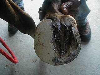 In this case, an extension to the hoof is fabricated with a compositec material and is attached to the side of the affected heel thus increasing the width of the hoof wall (Figure 5, 6). This serves two purposes. First, it causes the foal to break over straighter, improving the limb flight and resulting in a more uniform strike pattern. Second, the extension prevents further bending of the hoof wall, and adds support to the heel. As the hoof wall grows distally, it will in many cases follow the direction of the composite so that the wall has bends less. A large number of these foals will improve as they grow because, as the chest widens, the rotational deformity improves, changing the landing pattern. In this case, an extension to the hoof is fabricated with a compositec material and is attached to the side of the affected heel thus increasing the width of the hoof wall (Figure 5, 6). This serves two purposes. First, it causes the foal to break over straighter, improving the limb flight and resulting in a more uniform strike pattern. Second, the extension prevents further bending of the hoof wall, and adds support to the heel. As the hoof wall grows distally, it will in many cases follow the direction of the composite so that the wall has bends less. A large number of these foals will improve as they grow because, as the chest widens, the rotational deformity improves, changing the landing pattern. PROGNOSIS
The prognosis for this condition is good provided a skilled interested farrier is involved. It is also necessary to have a committed owner as resolution of these cases is slow and they often require ongoing maintenance. Theoretically, the prevention and treatment of lameness caused by inappropriate mediolateral orientation is simple but in practice it is often difficult to achieve. Being aware that sheared heels can predispose the horse to multiple hoof wall problems makes prevention imperative. When lameness is localized to a sheared heel or associated with sheared heels treatment becomes necessary, but sound horses with a sheared heel from whatever cause also will benefit from correction. Many times, improvement is all that can be achieved. Supplemental Readings 1. Turner TA: The use of hoof measurements for the objective assessment of hoof balance. Proc 38th Annu Conv Am Assoc Equine Practnr 1992; 389-395. 2. Moyer, W., Anderson, J. P.: Sheared heels: Diagnosis and treatment. J Am Vet Med Assoc 1975; 166:53. 3. Hickman, J. Humphrey, M. Hickman's Farriery. 2nd ed. 1988, London: J.A. Allen. 136-182, 195-227. 4. Snow V, Birdsall D. Specific parameters used to evaluate hoof balance and support. . Proc 36th Annu Conv Am Assoc Equine Practnr 1990; 299-311. 5. Balch O, White K and Butler D. How lameness is associated with selected aspects of hoof imbalance. Proc 39th Annu Conv Am Assoc Equine Practnr 1993. 6. Williams G, Deacon M, No Foot, No Horse. 1st ed. 1999, Buchingham: Kenilworth Press. 1-16, 30-48, 65-80, 99-111.
Figure Legends:
Figure1. Palmar view of sheared heel. Note disparity between medial and lateral heel length. Also note that the coronary band bulges abaxially to create a lip. Figure 2. Plantar view of sheared heel. Note the underrun heel. Figure 3. Shoe is too small with unequal branches. Note that the medial heel is displaced proximally.
Figure 4. Horse with sheared heels shod with full bar shoe. Note that there is a space between the affected heel and the shoe.
|