Equine Hoof Capsule Distortions: An OverviewReprinted with permission from the American Association of Equine Practitioners. |
|
Limb Conformation
When evaluating hoof capsule deformation, limb conformation should be considered. Abnormal limb conformation affects the landing pattern and stance phase of the stride. Few horses have ideal limb conformation and any change in conformation may lead to a change in the distribution of forces within the hoof capsule, leading to deformation. In the frontal plane, the forelimbs should be of equal length and size and bear equal weight.
| ||
|
Healthy Foot
A morphological description of what is considered to be a good or ideal foot can be used as a model not only to relate or compare a hoof capsule distortion but also as a template or guideline when formulating a farriery plan.11 Examination of the foot begins with the horse standing square on a firm, flat surface and the foot viewed from all sides. Following visual examination of the feet, the horse should be observed in motion, both going away from and toward the examiner, on a firm flat surface to note the landing pattern of the foot as it strikes the ground. Finally, the ground surface is examined with the foot off the ground. Additionally, small changes in the shape of the hoof capsule (such as the coronet and the digital cushion) may be further appreciated by careful palpation of the foot than by visual inspection.
Dorsal Aspect
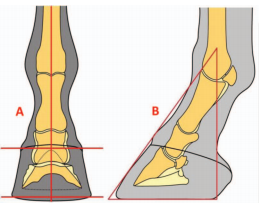
When the foot is viewed from the dorsal aspect, the ideal hoof should be approximately symmetrical.An imaginary line drawn between any two comparable points on the coronary band should be parallel to the ground. The medial wall should be the same height as the lateral wall, but because it is often slightly steeper, it may be slightly shorter. An imaginary line that bisects the third metacarpal should bisect a line drawn between any two comparable points on the coronary band or the ground surface of the hoof. Similarly, the hoof should be symmetrically related to the distal limb such that an imaginary line that bisects the third metacarpal bone bisects the pastern and the hoof, allowing for the slight asymmetry due to the different angles of the medial and lateral wall (Fig. 4A).1 When the foot is viewed from the dorsal aspect, the shape of the forefeet may be asymmetrical, with one hoof being narrower than the other (“mismatched feet”).On palpation, the coronary band of a healthy hoof should feel thick and spongy. There should be no evidence of a “ledge” or “trough” behind the proximal margin of the hoof capsule when palpated. A depression in the coronary band indicates that the distal phalanx has displaced within the hoof capsule, a finding that can be present in sound horses.12This palpable depression will generally be accompanied by a thin, flat sole, narrow frog, and contracted heels. The dorsal aspect of the coronary band should also be palpated for effusion of the DIP joint. This is often seen with horses that have a broken back hoof pastern axis (HPA) and synovitis of the DIP joint.
|
Lateral Aspect
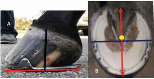
When viewed from the lateral aspect, the angle the dorsal hoof wall forms with the ground is variable and typically related to the conformation of the digit. The heel tubules of the hoof capsule should form an angle with the weight-bearing surface similar to the angle of the horn tubules in the toe region. Tradition has it that the angle of the wall at the heel should match that of the dorsal hoof wall at the toe;however, it varies and is generally a few degrees less. The length of the dorsal hoof wall is similarly variable, but is determined by the amount of sole depth present. There are two guidelines that relate the proportion of the foot to the rest of the distal limb. First, the foot pastern axis describes the relationship between the angles made by the dorsal hoof wall and the dorsal aspect of the pastern with the ground. Ideally, the dorsal hoof wall and the pastern form the same angle with the ground so that the angle between them is 180° and the axis is considered straight. Second, an imaginary line that bisects the third metacarpal should intersect the ground at the most palmar aspect of the ground surface of the hoof (Fig. 4B). The healthy coronary band should have a gentle, even slope from the toe to the heels and the hair should lie flat against the hoof capsule; hair projecting horizontally may indicate excessive forces on the associated hoof wall.1 The width of the growth rings below the coronet should be equal from toe to heel. A disparity in the width of the growth rings between the toe and the heels is indicative of nonuniform circulation of the coronary corium or excessive forces below, because wall growth is generally inversely related to load at the bearing border of the foot.
|
Palmar Aspect
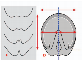
The heels are evaluated from the palmar aspect for their overall width and height. The heels frequently become narrower when the foot itself is narrow. The overall height of the heels is readily assessed from the lateral aspect but viewing from the palmar aspect is useful to compare the relative heights of the two heels when measuring from the hairline at the bulbs to the ground. For example, in the case of the sheared heel,one heel is displaced proximally relative to the other heel. Another example is mismatched feet where there is a marked disparity in heel height between feet. The contour of the junction of the heel bulbs with the skin can be evaluated relative to the width of the hoof wall at the heels and the thickness of the digital cushion (Fig. 4C).
Distal or Solar Aspect
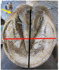
When viewed from the distal surface, the ground surface of the foot should be approximately as wide as it is long.1,2 The foot should be approximately symmetrical about the long axis of the frog; the lateral side of the sole frequently has a slightly greater surface area that corresponds with the difference in wall angles at the quarters described in the dorsal view. The width of the frog should be approximately 60–70% of its length.14 The ground surface of the heels should not project dorsal to the base of the frog, and the hoof wall at the heels and the frog should be on the same horizontal plane.Imaginary lines drawn across the most palmar weight-bearing surface of the heels and across the heel bulbs at the coronary band should be parallel and both lines should be perpendicular to the axis of the frog (Fig. 4D).1
The author further evaluates the solar surface of the hoof capsule by drawing a line across the widest part of the foot. This line forms a consistent landmark and is located just dorsal to the COR (of the DIP joint). Using this line as a starting point, there should be approximate proportions from this line to the perimeter of the toe and to the base of the frog.Hoof balance or a balanced foot has been used historically to describe the ideal hoof conformation.However, hoof balance has no consistent definition and remains a concept. The author prefers to use the term “proportional foot” to describe an acceptable foot conformation. This term can be used to access foot conformation from the lateral side as well as the ground surface of the foot by using the COR as the intersection between the proportions (Figs. 5A and 5B).
|
|
Long-Toe, Low-Heel Foot Conformation
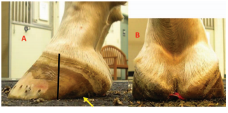
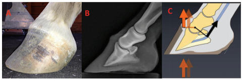
A low-heel conformation can readily occur with or without excessive toe length. This type of foot configuration is so common in equine practice, especially in Thoroughbred horses, that it is thought to be normal.15 A long-toe/low or underrun heel conformation (LT-LH) is defined as the angle of the heels being considerably less than the angle of the dorsal hoof wall. When this difference in angles is considerable, it is characterized by a broken back HPA where the angle of the dorsal hoof wall is lower than the angle of the dorsal pastern. It is often the result of leaving the heels to migrate dorsally when trimming, which allows them to grow forward and lose their angle. When evaluating the foot from the lateral aspect, there will be disproportionate distances on either side of the middle of the foot to the toe and to the heel. There may or may not be a flare in the dorsal hoof wall. The coronet will reveal an acute angle from the toe to the heel and the coronet at the heels will thicken and begin to form a “knob”-shaped appearance. The angulation of the horn tubules will decrease from toe to heel and may often be parallel with the ground at the heel (Fig. 6). The ground surface of the foot will again show a disproportionate distance from the widest part of the foot to the perimeter of the toe and to the base of the frog.The heels of the hoof capsule will have migrated dorsally while the soft tissue structures are located palmar to the end of the heels, and, in many cases,the frog is situated distal to the bearing border of the hoof wall. Interestingly, when observed in motion on a firm flat surface, a horse with LT-LH conformation may have a markedly heel-first landing due to the lack of ground surface in the palmar foot, the horse may land flat, or the horse may land toe first if they are experiencing discomfort in the palmar foot.
|
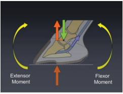
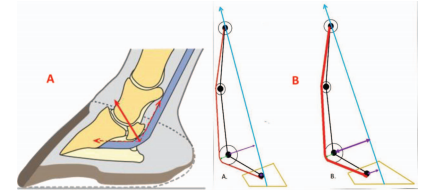
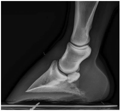
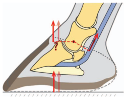
A low hoof angle results in dorsiflexion of the DIP joint, which concentrates weight bearing on the palmar section of the foot and increases strain on the DDFT. This excess load, in turn, may cause increased stresses on the navicular apparatus and the soft tissue structures associated with the palmar foot and the joints proximally16 (Figs. 7A and 7B).If low or underrun heels are allowed to progress,this condition can be readily observed both visually and radiographically; the point at which the angle that the hoof capsule or the distal phalanx forms with the ground is lower palmarly/plantarly than it is dorsally. A negative angle of the solar border of the distal phalanx, as noted radiographically, means that the soft tissue structures (frog, digital cushion)are underdeveloped or have decreased in mass, usually due to damage, or they have prolapsed palmarly which allows the distal phalanx to descend distally(Fig. 8). Biomechanically, it changes the angle of insertion of the DDFT on the distal phalanx, increases the peak force on the navicular bone bursa, and moves the GRF dorsally toward the toe(Fig. 9).4,5
|
|
Farriery
The treatment of low or underrun heels is difficult,and often the conformation of the heels can only be maintained rather than improved. Farriery seeks to reduce the length of the dorsal wall and redistribute the weight on the ground surface of the foot.The traditional farriery for low or underrun heels is to use an egg bar shoe to support the heels, often accompanied by some form of heel elevation to raise the angle of the heels and correct the broken back HPA. However, it is questionable whether “support”can be applied to compromised structures (heels) that no longer have the ability to accept weight, and the egg bar shoe may do little more that apply leverage to the palmar foot.
The ability to improve the soft tissue structures in the palmar foot and to produce new hoof wall grow that the heels may be limited. In the author’s experience, it appears that some form of structural framework is necessary to support renewed hoof wall growth at the heels. In the palmar foot, this “framework”seems to be the digital cushion, frog apparatus, and ungual cartilages, and when these structures are compromised, renewed hoof wall growth is poor or absent. Various techniques have been successful,depending on the amount and integrity of the structures present. When possible, the author has had success leaving the shoes off and allowing the horse to be barefoot for 30–60 days.17 This method is useful if the frog is located distal to the ground surface of the foot, as it will put the frog back on the same plane with the hoof wall. Otherwise, when a prolapsed frog is present, the shoe should be removed, excess horn trimmed from the frog, and the horse stood on a hard surface such as a rubber stall mat for 24–48 hours before the trim.
Foot preparation begins in all feet by visualizing the two basic landmarks on the ground surface of the foot: the widest part of the foot and the base of the frog. The palmar section of the foot is trimmed appropriately by using the widest part of the foot as a starting point, and the heels are trimmed to healthy horn when possible, making sure that all of the structures of the heel and the frog are on the same plane.The toe is shortened accordingly, again using the widest part of the foot as a guideline. Many horses with a LT-LH will have decreased sole depth, so, when necessary, the toe length can be reduced by using the nippers in a vertical plane across the toe (which preserves sole depth) rather than using them in the usual horizontal plane. Following the trim, the heels are assessed with regard to the structural integrity and the structural mass present.
|
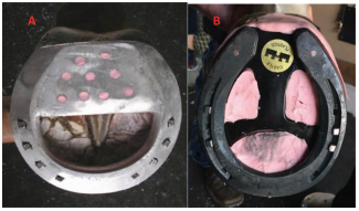
A shoe that attempts to place the load over the entire palmar foot complements the trim, not just on the hoof capsule at the heels (load sharing concept)and provides heel elevation to improve the HPA when possible. If the structures of the heel are intact and the hoof wall angle simply needs to be raised, an open aluminum wedge shoe or an open steel or aluminum flat shoe with a wedge pad can be used to achieve the desired heel angle. Depending on the severity that the heels are damaged or compromised, a load-sharing effect can be accomplished by using a straight-bar shoe with a pad or degree pad placed between the shoe and the foot. The author is reluctant to use a heart-bar show if the frog and digital cushion lack sufficient structural mass. An open-steel or aluminum shoe with a heel plate welded between the branches of the shoe or a plastic bar wedge placed between the shoe and the foot can also be used. Holes can be drilled in the heel plate or bar wedge and some form of silastic material placed underneath the plate or wedge to create a deformable interface to spread the weight bearing function over all the structures in the palmar foot. The same effect can be achieved by placing a “spider” plate between the shoe and the foot. Impression material is placed on the ground surface of the palmar foot, starting in the middle of the frog and extending palmarly as far as desired.The shoe and pad or plate are placed on the foot,and the heel of the shoe is pressed into the impression material, forming a slight wedge. Two nails are placed in the toe of the shoe and the foot is held off the ground until the impression material cures(Figs. 10A and 10B). Glue-on technology may be helpful to improve the structures in the heel but should not be applied long-term because damage to the hoof wall is thought to occur from the heat generated by the composite, and they have been shown to decrease expansion at the heels.18
|
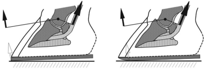
Farriery for low heel conformation is often based on trial and error and combinations of various methods but depends on assessing the structures present,the footing, the athletic pursuit of the horse, and client expectations. Any form of farriery for LT-LH should be accompanied by enhancing breakover.Moving breakover palmarly can be accomplished in a variety of ways, such as rolling or rockering the toe of the shoe or creating a rolled toe in the shoe by using a hand grinder where the breakover begins at the inner branch of the shoe. Moving breakover palmarly/plantarly decreases the moment applied to the DIP joint and appears to decrease the maximum tension in the DDFT, which occurs toward the end of the stance phase at the beginning of breakover (Fig.11).1–7
Upright or Clubfoot Conformation
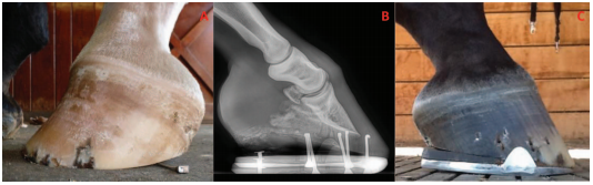
Flexural deformities have been reported as a cause of decreased athletic performance and chronic low grade lameness in the mature horse.19–22 A clubfoot is defined as an upright conformation of the foot associated with a flexural deformity of the DIP joint.19,21 It is characterized by a broken forward HPA, which is a reflection of a hoof capsule where the angle of the dorsal hoof wall is higher than the angle of the dorsal pastern. This broken forward HPA or flexural deformity is created by some degree of shortening of the musculotendinous unit (DDFT and associated muscle bellies), causing the DIP joint to be drawn into a flexed position. Biomechanically, as the tension in the DDFT increases, the COP moves dorsally in the toe (Figs. 12A–C).19,22
 |
| Fig. 12. A, Shows a clubfoot with a broken forward hoof pastern axis, the coronary band has lost the slope, and a flare in the dorsal hoof wall. B, A radiograph with a flexural deformity and C shows the biomechanics of a clubfoot; when the tension in the DDFT increases, the COP moves dorsally in the toe (Courtesy Dr. Andrew Parks). |
Examination from the lateral side generally reveals a broken forward HPA, the coronet assumes amore horizontal position, poor hoof wall consistency,a disparity of hoof wall growth with more growth at the heel than at the toe is generally present, and there will be some degree of a flare in the dorsal hoof wall. Looking at the ground surface of the foot, there will be disproportionate ground surface on either side of the widest part of the foot, with the palmar section showing less surface area, a thin sole, separations at the toe, and the frog generally receding due to excess hoof wall growth at the heels.Observing the horse in motion, depending on the severity of the flexural deformity, the horse will either have a toe first landing pattern or will land flat.
Farriery
High hoof angles with no or mild phalangeal misalignment can generally be improved by gradually trimming the heels in a tapered fashion from the apex of the frog palmarly to the heels. The solar surface of the foot dorsal to the frog should not be trimmed so all sole thickness is maintained. This increases the ground surface of the foot and attempts to reestablish weight bearing on the entire solar surface of the foot. The upright foot will often have a thin sole, so toe length is reduced accordingly from the outer dorsal hoof wall with an attempt to remove any concavity in the wall. A flat-steel or aluminum shoe fitted so it extends beyond the heels of the hoof capsule and breakover moved palmarly to the first nail hole to compensate for any increased tension in the DDFT created by lowering the heels is adequate. The polyurethane shoe (Polyflex®) provides another option to use with mild upright foot conformation due to its compliance of the flexible shoe with any DDFT tension and the mild heel elevation present in the shoe, which is enhanced by creating breakover in the shoe with a grinder (Robert Hunt, DVM, personal communication).
Farriery for a high hoof angle with concurrent phalangeal misalignment is a greater challenge.The object of farriery is to realign the distal phalanx within the hoof capsule, load the heels, and compensate for the shortening of the DDFT, all of which will improve the HPA. Therefore, farriery is directed at lowering the heels, but the amount to remove can be hard to determine. In mild to moderate clubfeet,the amount of heel to be removed can be estimated by placing the thick end of a 2° or 3° pad under the toe of the foot and allowing the horse to stand on it.If the horse does not resent the tension placed on the DDFT, the thickness of the degree pad can be removed in a tapered fashion starting at the widest part of the foot. The toe is shortened by backing up the dorsal hoof wall with a rasp. The trimmed foot is fitted with a shoe that has the breakover forged or ground into the shoe starting just dorsal to the apex of the frog and tapering toward the toe to further decrease the stresses on the DDFT.
A clubfoot with a marked flexural deformity should still have the heels trimmed in order to load the heels and unload the toe. However, heel elevation must be added to the shoe or incorporated into the shoe to compensate for the shortening of the musculotendinous unit. The necessity of adding heel elevation will also be evident if the horse had a toe-first landing pattern noted during the initial examination prior to the farriery. This can be determined following the trim by placing the trimmed foot on the ground palmar to the contralateral limb to observe for any space between the heels of the foot and the ground (Fig. 13A). The author uses a wedge shoe or places a wedge pad or a bar wedge between the heels of the foot and the shoe to compensate for the shortening of the tendon unit. If a wedge shoe is selected, silastic material should be placed over the solar surface between the branches of the shoe to support the sole which will be higher off the ground.This method allows the heels to be weight-bearing but at the same time decreases the stresses in the muscle tendon unit. Breakover is applied as described above (Figs. 13B and 13C). Severe flexural deformities that result in chronic lameness can be treated by performing an desmotomy of the accessory ligament of the DDFT combined with the appropriate farriery described above.9,22
 |
| Fig. 13. A, Clubfoot placed on the ground behind the contralateral forefoot to test for tendon shortening after heels are trimmed.Note the space under the heels. B, Radiograph of a clubfoot with a marked flexural deformity. Note the lucency in the palmar section of the image that denotes the receded frog. C, The same foot seen in B after being shod with heel elevation to compensate for the shortened muscle tendon unit and consequently be able to load the heels. Note the hoof pastern axis. |
|
Sheared Heels
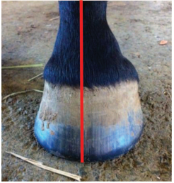
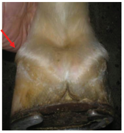
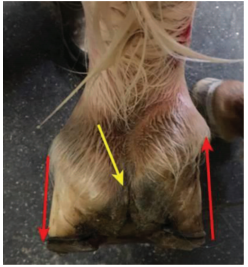
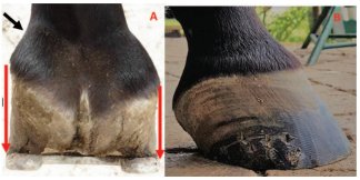
Sheared heels is a hoof capsule distortion resulting from displacement of one heel bulb proximally relative to the adjacent heel bulb.12,13,23 This disparity between the lateral and medial heel bulb is generally 0.5 cm or more. The displaced bulb is predominately seen on the medial side but can be seen on the lateral side. Sheared heels appear to develop as an adaption-distortion of the hoof capsule as a consequence of limb conformation that results in an abnormal strike and loading pattern of the foot on the ground. The author believes that when the adaptive ability of the hoof capsule is surpassed by the excessive load on one section of the foot, the ensuing type of hoof conformation can be a source of unilateral palmar foot pain and predisposes the foot to subsolar bruising, corns, quarter cracks, fracture of the bar, and deep fissures within the base of the frog (Fig. 14). This foot conformation is readily observed by standing behind the horse and noting the relative distances measured from the heel of the hoof capsule to the hairline at the bulbs of the heels and noting any difference between the lateral and medial heel. Observing the displaced heel/quarter from the side, the wall will be straighter due to increased load and may start to roll under; the coronet will be displaced proximally and assumes amore horizontal appearance, and there will be tightly packed growth rings below the coronet (Figs.15A and 15B). Biomechanically, the position of the coronary band is related to the balance between hoof wall growth at the coronary band and the rate of migration of the hoof wall distally (Andrew Parks,personal communication 2017).
|
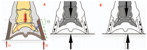 |
| Fig. 16. A, An illustration showing the balance between the growth of the hoof wall and its migration toward the ground, which is countered by the force on the hoof wall. B, Shows the COP moved to the overloaded side of the foot, which can be addressed by the appropriate farriery (Courtesy Dr. Andrew Parks). |
Farriery
Farriery is directed toward unloading the hoof wall and decreasing the forces on the displaced side of the foot. Because many horses with sheared heels will have a toe-out conformation, traditionally, farrier practices have advocated trimming the horse’s heels so the ground surface is lower on the opposite side from the side being displaced proximally. Intuitively, if the heel is longer on the displaced side(measured ground surface to hairline), it is reasonable to trim the displaced side. When possible, the author likes to remove the shoes and stand the horse on a hard surface for 24 hours prior to the farriery,as this allows the affected side of the foot to settle into a more acceptable conformation.
|
Farriery is initiated by removing the shoes and again observing the horse walking on a hard surface,noting the strike pattern of the foot. The author will use a double-trimming method in an attempt to improve and unload the distorted quarter/heel. As described previously, the trim begins with a line drawn across the widest part of the foot with a magic marker. The frog is trimmed to where it is pliable and the quarters and heels of the hoof capsule from the middle of the foot are rasped palmarly so the heels of the hoof capsule and the trimmed frog are on the same plane if possible. An attempt is made to create as much ground surface under the affected heel as possible, which will often result in more ground surface on the displaced side, which may make that side marginally lower than the other side of the foot. The toe and quarters are reduced appropriately so when the trim is completed, the surface area on either side of the line drawn or the widest part of the foot will approximate each other,resulting in a proportional foot (Fig. 17). Trimming the quarter/heel on the displaced side of the foot is logical, as it is the taller heel and it increases the ground surface of the foot on that side. Following the trim, the horse is again walked on a hard surface and some improvement in the landing pattern is generally noted.
 |
| Fig. 18. A, A steel straight bar shoe with the nailing pattern for a sheared heel. B, Shows the platform created under the heels by the bar shoe with the medial heel unloaded. C, Shows the space created under the displaced heel at breakover. Note the displaced coronet and the hoof wall defect present. |
If the displacement is significant, the author’s choice is a wide, web steel, straight-bar shoe fitted symmetrically to the trimmed foot (Fig. 18A). Bar shoes effectively increase the surface area of the foot, allow the palmar/plantar section of the foot to be unloaded, and decrease the independent vertical movement at the bulbs of the heels. If the displacement of the quarter/heel is marginal, an open-heel shoe can be used, but the trim remains the same.Before applying the shoe, a second trim is performed under the proximally displaced quarter heel, which goes from 0 mm at the ipsilateral toe (e.g., inside toe for medial sheared heel) to an average of 7 mm at the affected heel. The amount of heel that can betaken off in the second trim depends on the sole depth at the seat of corn and on the severity of the proximal displacement of the coronary band at the sheared heel. The amount of horn, under the sheared heel, which can be taken off with this second trim, ideally corresponds to the difference in length/height between the two heels. Lowering the hoof wall at the quarter/heel will create a space between the shoe and the hoof wall on the displaced side of the hoof (Figs. 18B and 18C). This improves the landing pattern, unloads the affected heel, and allows the heel bulb to settle down and assume amore acceptable position. Feet with a low palmar/plantar angle rarely have enough sole depth under the affected heel for the second trim; in these cases,the rest of the hoof wall can be raised with a full leather or synthetic pad and impression material.Impression material is placed in the palmar section of the foot from the apex of the frog palmarly except under the displaced heel/frog sulci where the second trim was performed. After the shoe is attached to the foot, the affected heel will rapidly descend onto the shoe, making the original space created by the second trim between the hoof wall and the shoe disappear. As most horses with a sheared heel have a predisposing limb conformation (e.g., a rotational deformity), these feet have a tendency to continue to deform the affected heel proximally and the double trim method usually has to be applied to some degree at each consecutive shoeing. Horses with this type of hoof conformation should be reset at 4–6-week intervals.
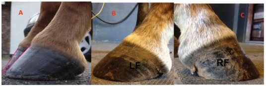 |
| Fig. 19. A, Shows the difference in dorsal hoof wall angles in a pair of mismatched forefeet. B and C, A side view of a pair of mismatched forefeet where the LF has a low angle while the RF has an upright angle with a mild broken forward hoof pastern axis. |
Mismatched Feet
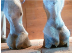
The management of mismatched hoof angles remains a controversial subject for both the farrier and veterinarian. Mismatched feet could be defined as forefeet conformation that have a high or upright hoof angle on one foot and a low hoof capsule angle on the contra lateral foot (Figs. 19A–C).21,24The difference between the forefeet could range from a high hoof angle with a straight HPA to a clubfoot with a flexural deformity and an overloaded low heel on the contra lateral limb. The mass, integrity, and difference in heel height will be the contributing factors to the mismatched dorsal hoof angles (Fig.20). Limb length disparity has been suggested as a cause for mismatched feet, although it has not been scientifically proven. Mismatched feet may contribute to poor performance, subtle lameness, and a shortened anterior phase of the stride on the upright foot. Traditional farriery seeks to elevate the heel on the low foot and therefore match the forefeet.However, this practice should be discouraged and treatment should be based on farriery principles to improve the structures and function of the individual foot.
|
Observation again begins by standing the horse on a firm flat surface. Looking from the front, the hoof with the upright hoof angle will be narrower than the hoof with the low hoof angle.The HPA should be evaluated from the side with the third metacarpal bone always perpendicular with the ground. It should be determined whether the foot with the high hoof angle has a straight or broken forward HPA and whether the foot with the low angle has a straight or broken backward HPA (Figs. 21A and 21B). If the foot with the high hoof angle has a broken forward HPA, it should be considered to have a flexural deformity or a clubfoot (Fig. 21C).Looking at the heels from behind, the integrity of the structures in this area should be evaluated and the difference in the height of the heels should be noted. It is also important to note whether the frog is recessed between the heels of the hoof wall on the foot with the high heel and whether the frog is prolapsed distal or palmar to the hoof wall on the hoof with the low hoof angle. Looking at the bottom of the foot, it is helpful to visualize a line drawn across the widest part of the foot and look at the proportions of the foot on either side of the line. Again, it is important to note if the frog is distal to the hoof wall or whether the frog is recessed below the heels of the hoof wall. The frog being recessed causes impaired function in the palmar section of the foot and places the entire load on the hoof wall.The horse should be evaluated in motion at both the walk and the trot. First and foremost, it is important to rule out any lameness. It is especially important to evaluate the landing pattern of the forefeet, as the foot with the high hoof angle will often land toe first rather than flat. Lastly, the horse should be trotted to note whether the horse has a shortened stride on the high-heeled foot.It must be remembered that a shortened stride on one limb will cause the opposite foot to be on the ground longer, which, over time, may create further damage to the heel structures of the foot, with the low heel resulting in a flat “panned out” low foot angle.
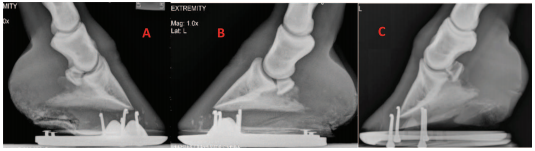 |
| Fig. 21. Mismatched feet. A, Upright foot with a straight HPA. B, A foot with a low hoof angle with a broken back HPA. Note the small shoes on both A and B. C, An upright foot with a broken forward HPA, defined as a flexural deformity or clubfoot. |
Farriery
Horses with a disparity between dorsal hoof wall angles will generally have a straight HPA and the hoof wall growth below the coronet from the toe to the heel will be even. In this case, the author suggests using good farriery principles to apply the appropriate trim and shoe for each foot on an individual basis. These basic farriery principles include the following:
Managing horses with mismatched feet where one foot has a high hoof angle with a flexural deformity or a clubfoot becomes more complex. This type of case will often present with a shortened stride on the limb with the upright foot. Low or compromised heel structures may be noted on the opposite foot from overloading the heel on that side due to the shortened stride placing excess weight on that foot over time. Managing these horses can be difficult and the proper shoeing protocol may not be inherently obvious. Again, it must be emphasized that each foot should be approached on an individual basis (Figs. 22A, 22B and 22C). It is common to see horses with mismatched feet shod with two different size shoes; often a smaller shoe is used on the upright foot. This practice should be discouraged, as the ground surface on both forefeet should be the same.
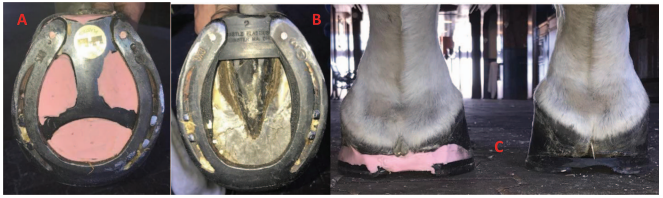 |
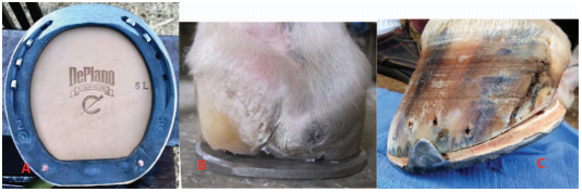
| Fig. 22. Mismatched forefeet shod according to their individual conformation. A, A low heel with a spider plate and impression material. B, A high heel with a wedge insert for heel elevation. C, A palmar view of A and B, showing the appropriate farriery for the foot conformation |
Farriery for the clubfoot has been discussed previously in this paper. When approaching the foot with the low angle, the clinician is often inclined to wedge up the heels to improve the HPA. However,this will place more stress on the already compromised heel structures. Although the HPA will appear improved immediately following the shoeing,the long-term effect is exacerbation of the low angle,further crushing of the heels, and prolapse of the frog below the ground surface of the foot. Alternatively, the heels should be trimmed back to the widest point of the frog if possible, or an attempt should be made to get the hoof wall at the heels and the frog on the same plane. As much toe length as possible should be reduced, generally using the dorsal hoof wall, as there is usually decreased sole thickness in the foot with a low heel. It should be emphasized that when possible, it is extremely important to obtain good quality radiographs prior to trimming to determine the amount of heel and especially sole that can be removed. Breakover, such as a rolled toe or a rocker toe if there is adequate sole thickness to allow it to be trimmed into the foot, is very beneficial, as it will further decrease the leverage at the toe on the low-angle hoof. The COP on a low-angle foot is further palmar than that of the upright or normal hoof, therefore, the shoeing protocol is directed at moving the COP away from the overloaded heels. Additionally, redistributing the load or load sharing with the weight-bearing structures of the low-angle foot may help to decrease the forces directed to the heels. This can be accomplished with impression material, a pour-in pad, and a spider plate or a heel plate added to the palmar aspect of the shoe.
Conclusion
The clinical examination of the equine foot has been well described and is generally performed in lameness cases.14 Evaluation of the hoof capsule during the lameness examination will not only provide additional information as to the etiology and treatment of the lameness but will also serve as a guideline to apply therapeutic farriery and other preventive measures to maintain a healthy hoof. The morphology of the hoof capsule reveals deformation and changes in growth that occur following increased or reduced force. The relationship between the limb and the foot indicate conformations that predispose the foot to abnormal weight bearing. Inversely, using the abnormal distribution of forces and the subsequent hoof capsule distortion as a template,appropriate farriery or therapeutic farriery will form at least part of the treatment plan. Here, it is essential for the clinician to be familiar with the biomechanics of the foot and how these forces can be altered to change the distribution of forces or the focal stresses on a given section of the foot.
Declaration of Ethics
The Author has adhered to the Principles of Veterinary Medical Ethics of the AVMA.
Conflict of Interest
The Author has no conflicts of interest