Equine Thrush: A Closer LookReprinted with permission from the American Farriers Journal. |
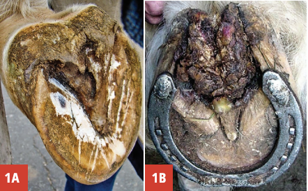 |
| Figure 1A&1B: Figure 1a is thrush. Note the deterioration of the frog and the recession below ground surface of the foot. Figure 1b is severe canker. Note the proliferation of the diseased horn. |
Etiology Of Thrush
The overall health of the frog is an important component of the etiology of thrush. A healthy, well-formed frog is broad, firm and deforms with thumb pressure. The frog should fill the space between the heels of the hoof capsule and, when measured at its base (the widest part), the frog width should be at least 70% of its length.4 A large, healthy frog acts as an expansion joint that holds the heels apart, shares the load-bearing function with the other structures of the palmar/plantar hoof and helps to absorb concussion. Under normal conditions, weight bearing stimulates the normal physiology of the frog, ensuring its continued health. A well-shaped frog that fills the space between the heels also promotes a natural self-cleaning mechanism such that when the foot is loaded, the frog expands, expelling any accumulated dirt or debris from the frog sulci.
An unhealthy frog is markedly smaller in size and recessed below the level of the ground surface of the hoof, thus creating a void in which debris can accumulate. This can occur for a variety of reasons, including many of the recognized hoof capsule distortions such as the low heel, clubfoot or sheared heels along with inappropriate farriery practices. Other contributing factors for thrush include wet, unhygienic stable conditions, especially when horses continually stand in urine and manure-soiled bedding; neglecting daily routine foot care; and lack of exercise.1,2,3,5 The fundamental problem with palmar/plantar conformation usually involves the frog not being near or on the same plane as the heels, thus not contacting with the ground, which reduces the stimulation from the ground, causing the frog to atrophy.
As the frog recedes within the hoof capsule, the frog loses its “self-cleaning mechanism,” which allows material (dirt, manure, etc.) to accumulate over the frog, creating excessive pressure. Over time, this constant pressure on a compromised frog leads to increased deterioration and atrophy. Weakened by the reduced protective horn of the epidermis, the frog tissue becomes susceptible to penetration by bacteria and, consequently, development of the disease. Furthermore, the accumulation of debris over the frog creates an environment conducive to bacterial growth, especially for anaerobic bacteria. The recessed frog loses its ability to act as an expansion joint and the heels begin to contract. The diseased frog can no longer share in supporting the horse’s weight, which shifts most of the load-bearing function solely onto the heels of the hoof capsule.
No specific organism has been identified as the sole cause of thrush. Two anaerobic bacteria species, Bacteroides sp. and Fusobacterium necrophorum are opportunistic organisms found in the soil and are commonly isolated from the bottom of the horse’s foot. On the positive side, when the necrotic tissue of the frog is debrided and the conformation of the structures in the palmar/plantar foot are improved, the anaerobic conditions will be improved and bacterial growth will be decreased. Furthermore, if antimicrobials are required, these organisms are quite susceptible to most topical preparations.
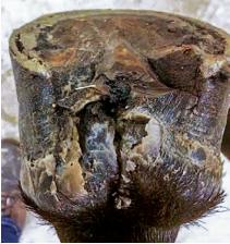 |
| Figure 2: This picture shows an untrimmed foot with thrush, the frog recess below the ground surface of the foot, sheared heels and a fissure present in the central sulci of the frog, extending to the hairline. |
Clinical Signs And Diagnosis Of Thrush
The diagnosis of thrush is usually straightforward and based on examination of the frog and surrounding structures. There is usually an increased amount of moisture on the bottom of the foot and a black exudate in the sulci of the frog. This exudate, which varies in quantity, usually has an offensive odor.2,5 The affected sulci of the frog are often deeper than usual and may extend into the sensitive tissues of the foot, causing them to be painful.
The frog will generally be small, recessed within the hoof capsule, lack a firm consistency and have a necrotic appearance. There may be a fissure present in the central sulci extending from the base of the frog to the hairline at the bulbs of the heels (Figure 2). The frog may also be undermined and areas of necrotic horn can often be detached from the underlying tissue.4 Lameness is present in severe cases that involve the dermis, and swelling of the distal limb may be seen.
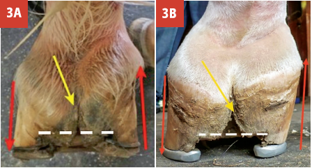 |
| Figure 3A&3B: Figures 3A and 3B both present foot conformation showing the frog receded and asymmetric foot landing, leading to sheared heels. The difference in force on each heel is exerted in opposite directions and results in the fissure in the base of the frog. Figure 3b is a severe clubfoot combined with a sheared heel. Note the loss of frog mass between the heels. |
Fissure In The Central Sulci Of The Frog
There will often be a fissure in the central sulcus of the frog that may be associated with thrush. The author believes the fissure is a primary structural defect in the soft tissue structures in the palmar/ plantar section of the foot that secondarily becomes infected, leading to thrush. The fissure is often seen in horses with a hoof capsule distortion (such as low heels with instability and sheared heels), a recessed frog, compromised soft tissue structures and an abnormal strike pattern.
The asymmetrical landing pattern results in an unequal force placed on each heel, causing the heels to move in opposite directions. This movement in opposite directions causes a fissure to form in the weakest part of the soft tissue, which is the central sulcus of the frog (Figures 3A and 3B). Significant movement in the fissure can often be demonstrated by moving the heel bulb in opposite directions.
Treatment Of Thrush
Farriery. Treatment for thrush begins with improving the hoof capsule, the conformation of the palmar/plantar foot and the health of the frog. Thrush will not be resolved as long as the frog remains recessed below the ground surface of the foot. The amount of improvement in the frog is correlated with the amount of chronic damage, especially involving the frog dermis. If the horse can be given time off from work, the shoes should be removed, the heels trimmed to the same plane as the frog, the perimeter of the hoof wall rounded and the horse left barefoot.
When barefoot, load is shared by all the structures of the palmar foot and the integrity of structures improves rapidly and it appears to promote regrowth.6 Treatment of the thrush as described below is performed daily.
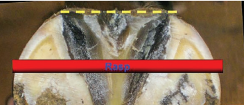 |
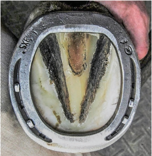
| Figure 4: Palmar section of foot showing heels trimmed to the base of the frog with the heels of the hoof capsule and the frog on the same plane. |
If shod, the basic trim begins by drawing a line across the widest part of the foot and trimming the heels of the hoof capsule such that they are on the same plane as the frog (Figure 4). This can generally be accomplished in most cases. The toe length is reduced dorsally to the line drawn across the foot, resulting in approximate proportions on either side of the line.6,7 If significant heel is removed from an upright or club foot, heel elevation may be necessary to accommodate the resultant stresses placed on the deep digital flexor tendon. When a fissure is present in the central frog sulcus, it should be stabilized to decrease the vertical movement of the heels that will inhibit healing. This can readily be accomplished by applying a straight bar shoe (Figure 5)
 |
| Figure 5: A straight bar shoe used to stabilize the hoof capsule and the vertical movement of the heels. |
Medical. Any detached areas of necrotic horn on the surface of the frog should be debrided. The frog is cleaned initially by soaking the foot in a saturated (add salt until it no longer dissolves) solution of Epsom salts (MgSO4). Topical antiseptics/ astringents including 2% iodine, merthiolate, chlorohexidine and various copper sulfate solutions are applied on a daily basis. Topical antibiotic ointments/solutions appear to be unnecessary and caustic preparations that contain phenol, formalin or formaldehyde should be discouraged.
Daily aftercare. Horses should have their feet cleaned and the frogs brushed vigorously with a stiff brush, after which an astringent is applied on a daily basis.1,2,3 If there is a fissure present in the central sulcus, the area above the fissure should be flooded with the antiseptic/astringent solution to allow penetration into the fissure, and then packed with a folded gauze pad. The horse should be kept in a dry, clean stall and bedded on wood shavings or sawdust. Turn-out should be in a dry paddock. Exercise should be encouraged to improve the foot’s normal physiology.
Repeated trimming of any detached horn on the frog may be required until the infection is controlled. Routine farriery should be scheduled every 4 to 5 weeks, paying close attention to the trim, the foot conformation, the frog and the integrity of the soft tissue structures of the foot.
Conclusion
There is a plethora of commercial products purported to treat and resolve thrush. However, unless the health and structural integrity of the frog is improved, these products are of limited value. It is rare to see thrush in a foot with a healthy frog and support structures in the palmar/ plantar foot.
A review of the literature published on the cause and treatment of thrush does not consider the role of farriery in the etiology of this disease process. Acquired hoof capsule distortions such as a low heel, club foot and sheared heels all affect the foot’s palmar/ plantar soft tissue structures and the frog’s health. Therefore, the health of the frog can’t be improved and, consequently, the thrush can’t be resolved without using appropriate farriery. Remembering that prevention is always superior to treatment, emphasis should be placed on good farriery as the primary means for prevention of this disease.
REFERENCES