|
How to Apply the Appropriate Farriery Principles to the Horse with Low Heels in the Hind Feet Reprinted with permission from the American Association of Equine Practitioners. |
|
Accepted farriery for low heels in the hind feet has been to provide heel elevation regardless of the foot conformation or integrity of the hoof capsule. Long egg bar shoes with wedge pads are generally used for this purpose. This is also the farriery that is generally prescribed for lameness localized to the hock or stifle, yet there is no documentation that confirms that heel elevation exerts any significant influence on any section of the hind limb above the distal interphalangeal (DIP) joint.9 Furthermore, heel elevation will tend to exaggerate a heel first landing and thus increase the pressure exerted on the hind feet that have existing low or underrun heels, which appears to compromise the structures of the hoof capsule further and lead to additional lameness problems.
Hind Limb Movement
Before discussing farriery for the hind foot, a brief description of hind limb movement is essential to understand the propulsionary function of the hind limbs.10 Protraction, the foot lifting from the ground, begins with the flexion of the hip, stifle, and hock; this action overcomes the inertia of the hind limb so minimal muscular work is done. The hip joint is flexed by the iliopsoas muscle, the stifle is flexed by the biceps femoris muscle, and the hock flexes as a result of the reciprocal apparatus. The fetlock also automatically flexes because of the tendinous nature of the superficial digital flexor tendon that travels from the hock to the pastern.
Retraction is accomplished by the middle gluteal muscle, which is attached to the femur above the center of rotation (the hip joint).10 As the middle gluteal muscle contracts, it rotates the whole limb backward. The hamstring muscle group (semitendinosus, semimembranosus, and bicep femoris) runs behind the center of rotation, so they form the second part of retraction. This movement is accompanied by contraction of the quadriceps muscles, which extends the stifle and, consequently, the hock and fetlock.
The stance phase of the stride starts with foot impact. Initially, the vertical impact stops but horizontal movement continues, which means the hoof slides forward before full weight bearing takes place. At full weight bearing, the retractor muscles continue to be engaged and drive the horse’s body forward with the foot fixed to the ground. An important part of the forward propulsion provided by the hind limb is the opening or extension of the hock joints during the second half of the stance phase.
At landing, the forefeet have the greatest vertical force and also experience peak DIP joint flexion, while the hind feet have a greater vertical force during the stance phase, with peak DIP joint flexion following horizontal movement; this would imply the forelimb is a pendulum that is swinging and absorbing force, while the hind limb is grabbing and generating force.10
A Good Hind Foot
The authors are reluctant to use the term “normal” to describe hind foot conformation. The terms good, ideal, or functional may be more appropriate, as foot conformation with its inherent shape is associated with many variables such as genetics, breed, limb conformation, and farriery. The hind foot has a steep hoof angle, the shape is narrow or conical, the toe is pointed, and the sole has a much deeper concavity when compared with those of the front foot. In the well-conformed hind foot, the lateral wall will have some degree of flare while the medial wall will be straight, the amount of which will be conformationally dependent. The shape of the hind feet is an indication of being designed for primary propulsion/traction and secondary weight bearing. The front foot is generally as wide as it is long, whereas the hind foot is longer than it is wide.
 |
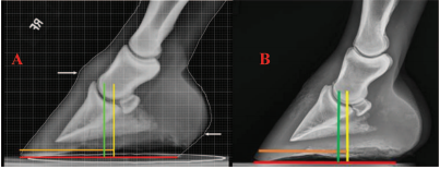
| Fig. 2. A, Lateral view of a hind foot with good conformation. Black line is the hoof pastern axis, red line is the middle of the foot, yellow line is the proportions of foot on either side of the middle of the foot, and green line is the appropriate length of a hind shoe. B, Solar view of a hind foot. Red lines are the widest part of foot and the proportions of ground surface on either side, and the yellow line is the base of the frog. Note the widest part of the foot is located further plantarly compared to the forefoot. |
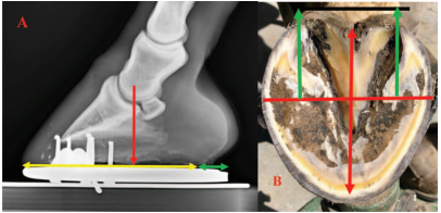
Looking from the side, a good hind foot will have a straight hoof pastern axis, even growth rings distal to the coronet from the toe to the heel, and approximate proportions on either side of the widest part of the foot (Fig. 2A). Looking at the solar surface of the foot, a line drawn across the widest part of the foot should divide the foot into approximate proportions on either side of the line. Considering the shape of the foot, it appears that the widest part of the foot is located further plantar in the hind foot when compared with that of the front foot (Fig. 2B). As the widest part of the foot is generally located 5–10-mm dorsal to center of rotation (COR), this difference between the fore and hind feet could be verified using lateral radiographs.a The first author did a small extempore study on a limited number of hind foot lateral radiographs supplied by two large equine referral centersb,c that were considered to be representative of acceptable conformation for a hind foot. The radiographs were measured and the proportions on either side of the COR were compared with forefoot lateral radiographs. The proportions generally found on a front foot with good conformation are 53–57% dorsal to the COR and 43–47% palmar to the COR.c,11 In all radiographs, the COR was found to be further plantar in the hind foot, which significantly decreased the ground surface of the foot plantar to the COR. Furthermore, in all radiographs, the digital alignment was not completely straight, as the middle phalanx was mildly displaced in a distal plantar direction relative to the distal phalanx (Fig. 3).
 |
| Fig. 3. A, Acceptable conformation for a forefoot (Courtesy Dr. Andrew Parks). B, Acceptable conformation for a hind foot (Courtesy Dr. Kurt Selberg). Yellow line is COR, green line is widest part of the foot, brown line is distance from COR to dorsal hoof wall, and red line is ground surface on the solar surface of the foot. |
Clinical Examination
Observations
|
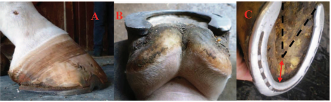 |
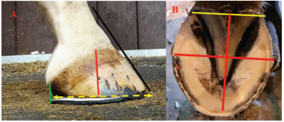
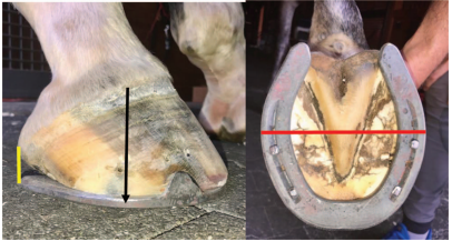
| Fig. 5. A, Lateral view of a moderate low heel “bull nose” conformation of a hind foot. Note the disparity in growth rings and the acute angle of the coronet. B, Plantar view shows the prolapsed frog as well as the incline of the frog. C, Solar view shows the incline of the frog in a dorsal cranial direction (black lines), and the marked concavity of the sole (red arrow). |
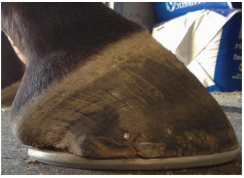
The Low Heel “Bull Nose” Foot Conformation
This abnormal conformation of the hind feet is easy to recognize. When looking at the limb from the side, the digit will show a broken back hoof pastern axis. The slope of the coronary band from the toe to the heel will have an acute angle of 40–45°, and the coronet will bend distally at the heel to become almost vertical. The bulbs of the heels will be prolapsed plantar to the heels of the hoof capsule and will form a “knob”-shaped appearance that can be seen lying against the shoe. The hair on the coronet at the heels may project horizontally rather than lying flat against the hoof due to excessive load on the associated hoof wall. There will be a disparity in the growth rings below the coronet from the toe to the heel, with the growth rings wide apart at the toe and then tightly packed at the heel. The dorsal hoof wall will assume a “bull nose” appearance (Fig. 5A). Looking at the foot from behind, the frog will be large and bulbous from the constant stimulation with the ground, a ledge will form in the frog from bearing weight, and it will be situated well below the hoof wall with the bulk of the frog now located between the two branches of the shoe (Fig. 5B). The solar surface of the foot will show an inclined plane of the entire frog from the base to the apex in a dorsal cranial direction toward the coronet. This inclined plane or angle will match the angle of the solar border of the distal phalanx in the hoof capsule. The toe area on the solar surface of the foot will show a deep or exaggerated concavity between the apex of the frog and the inner branch of the shoe instead of a steep, yet smooth transition of the sole from the frog to the sole wall junction. There will usually be a palpable “trough” located just dorsal to the apex of the frog (Fig. 5C). Upon removing the shoe, the end of the heel of the hoof capsule is located well forward from the base of the frog and the horn tubules will be parallel with the ground. The hoof wall at the heel will be thin, the bars may be damaged or missing, and the angle of the sole will be absent. Lightly paring the area adjacent to the hoof wall at the end of the heel with a hoof knife will often show moderate to severe hemorrhage from the pressure of the damaged hoof capsule against the shoe. When the foot is placed on the ground, total weight bearing will be placed on the frog, which is located distal to the ground surface of the hoof capsule, and many horses will be reluctant to stand on it when the opposing limb is lifted off the ground. As noted previously, hoof testers placed on either side of the heel at the angle of the sole will often elicit a painful response and the structures will deform (Figs. 6A, 6B).
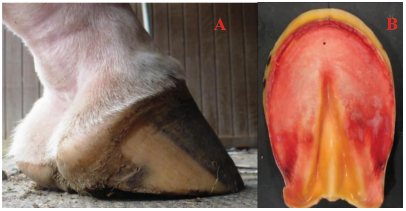 |
| Fig. 6. A, Hind foot from Figure 5 with shoe removed. Note the frog located below ground surface of the foot and the horse standing on the frog rather than the hoof capsule. B, A necropsy specimen from a horse with severe low heels in the hind foot. Note the hemorrhage in the sole. (Courtesy Michael Savoldi). |
|
Radiographs
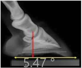
A lateral radiograph of the hind foot will show a broken back hoof pastern axis, with the middle phalanx (P2) being displaced plantar and distal relative to the distal phalanx (P3) during weight bearing. This places excessive stresses on the plantar section of the hoof capsule. The COR is located further plantarly with this abnormal foot conformation, thus decreasing the ground surface in the plantar section of the foot. The soft tissue structures (frog, digital cushion) in the plantar section of the foot have prolapsed plantarly to the shoe, forming a “knob”-shaped appearance. The angle of the solar border of the distal phalanx at the heels is lower than the dorsal margin of the distal phalanx (i.e., a negative plantar angle). The sole depth below the dorsal margin of the distal phalanx is markedly increased relative to the sole depth at the heel, and the perimeter of the dorsal margin of the distal phalanx can be seen migrating toward the dorsal hoof wall. The displacement of the distal phalanx results in the “bull nose” appearance of the dorsal hoof wall (Fig. 7).
Farriery
The amount of improvement that can be achieved with the appropriate farriery will obviously be proportional to the severity or the amount of distortion present. Damage to the plantar section of the hind feet is easier to improve than in the forefeet, possibly due to the anatomy and the difference of the load encountered on the hind limbs. The initial goal of the farriery is to make the plantar section of the foot “load sharing” such that the hoof wall and the frog are on the same plane. The first step of the farriery process will be to address the frog being located below the hoof wall. This will depend on the severity; if mild, the horse could have its shoes removed the day before being shod and housed on a firm surface, or if more severe, allowed to go without hind shoes for 3–5 days, which can be very effective. To begin the trim, the shoes are removed and the toe length is reduced from quarter to quarter according to the sole depth. Caution is advised when decreasing the toe length in this type of foot conformation, as the amount of sole depth noted on the radiograph or determined from the incline of the frog can be misleading. The dorsal margin of the bone migrates dorsally and therefore stretches the width of the dermis, a change which may not be recognized on the radiograph (Fig. 8A). Therefore, aggressive trimming at the toe will often result in seepage of blood at the sole wall junction as the dermal tissue is being encroached. It may be prudent to reduce the amount of sole depth gradually over two shoeing intervals. After the hoof wall is removed on the solar surface of the foot, additional horn is removed from the outer hoof wall to create even or uniform hoof wall thickness from quarter to quarter. The horse is then placed on a firm surface, which places pressure on the frog that quickly assumes the same plane as the heels on either side.
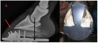 |
| Fig. 8. A, Lateral radiograph of a low heel “bull nose” foot conformation. Note the limited ground surface on the plantar side of the COR and the leverage created on the dorsal side of the COR. Lucent area under the red arrow shows the dermis being stretched as the margin of the distal phalanx migrates dorsally. B, Illustrates a frog plate created from a degree pad placed over the frog. |
If the frog prolapse is severe, the approach can be modified and the time frame shortened. The hind shoes are removed a day or two before the horse is due to be shod, and the foot is trimmed as described above. A frog plate is cut from a degree pad to match the frog, and the front of the pad is left intact to form a half moon design. The pad is attached to the foot with two small nails at the toe, and the foot is wrapped in a medicated poulticed that has been soaked in hot water and then secured to the foot with brown gauze and elastic tape (Fig. 8B). The horse is placed in a stall on a firm surface for 24–48 hours. When the wedge pad is removed, the frog will be compressed between the heels, forming a flat even plane which includes the frog and both heels. The horse will then be ready to have shoes applied, paying strict attention to the trimmed foot.
 |
| Fig. 9. A, Lateral radiograph of a hind foot that shows the concept of increasing the ground surface plantar to the COR using a shoe. (Courtesy Dr. Sarah Puchalski). B, Trimmed foot shows how the length of shoe (green arrows) will create approximate proportions on either side of COR foot. |
A line is now drawn across the widest part of the foot. Any additional horn at the heels can be removed using the rasp in a horizontal direction across the heels and frog so the hoof wall approaches the base of the frog to create as much ground surface as possible. Care must be taken to keep the frog and both heels in the same plane. When the hoof wall and the frog are on the same plane, the load is shared across the plantar section of the foot. The foot is now ready to have a sturdy steel shoe fitted and applied. The first author fit shoes to the hind feet in a similar manner to the front feet by using the line drawn across the widest part of the foot placed in the middle of the shoe. However, in the hind feet, the widest part of the foot will be located further plantar than the forefeet; therefore, additional shoe length is required to create the desired proportions on either side of the COR (Figs. 9–11). Looking at the shod hind foot from the side, the branch of the shoe should extend to or close to the point that coincides with a vertical line dropped from the hairline at the bulb of the heel. If the branch of the shoe extends beyond the vertical line or if the foot is not trimmed appropriately, the length of the shoe will create excessive leverage on the heels. In order to keep the frog and hoof wall on the same plane for the first shoeing interval or if mild heel elevation is necessary, a metal or aluminum heel plate or a 2° leather wedge can be placed under the shoe at the heels as long as the shoe is fitted in the manner described above. This will concentrate the load across the frog and heels rather than behind the heels, which is the case with a long shoe or trailers. The plate or wedge will also prevent the frog from descending toward the ground between the branches of the shoe. This is usually a temporary measure and it can be discontinued once the heels have stabilized.
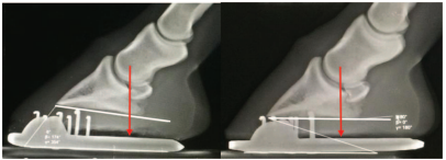 |
| Fig. 10. Lateral radiograph of a low heel “bull nose” conformation of the hind foot before and after farriery. Note the difference in the foot conformation after the appropriate trim and a size larger shoe. Again, note the increased ground surface plantar to the COR. (Courtesy Dr. Hans Castelijns). |
 |
| Fig. 11. The goal of hind foot farriery is to create a foot that has approximate proportions or ground surface on either side of the widest part of the foot. Note the branches of the shoe used to increase ground surface in the plantar section of the foot. |
Summary
The low heel “bull nose” foot conformation of the hind feet is often overlooked as a cause of poor performance or lameness in the hind limbs. This abnormal foot conformation may play a dual role in hind limb soundness. Firstly, the overload and damage to the plantar section of the foot can cause pain and a subtle bilateral lameness. Secondly, pain and/or the altered biomechanics of the foot will cause changes in hind limb movement. The changes appear to affect flexion of various joints, strain on ligaments, hoof and limb flight, hoof landing/loading, and muscle tension. Veterinarians and farriers frequently use specialty shoes or modify existing shoes in an attempt to relieve pain and therefore improve overall hind limb mechanics. However, it is unclear whether these modifications have an effect on hind limb biomechanics, as there is no research or current literature to support these modifications. It is the authors’ opinion that farriery begins with the appropriate trim, with the correct size/placement of the shoe with any subsequent modifications being secondary. The appropriate trim improves many hind limb issues by providing increased ground surface in the plantar section of the foot combined with a shoe of sufficient length so that the transition from loading to propulsion minimizes dorsiflexion of the fetlock and hock. In turn, this allows the toe to push off, elevating the limb into the swing phase, resulting in a smoother transition of force from the hind limb through the sacroiliac and lumber regions.
The incidence of this low heel “bull nose” hind foot conformation in performance horses has reached epidemic proportions in recent years. There are a myriad of theories/thoughts regarding how to trim the palmar/plantar section of the foot; however, the authors feel that inappropriate trimming of the heels decreases the ground surface in the plantar section of the hind foot and the application of shoes that are too small may be the inciting cause of this foot confrontation.
Treatment to address this foot conformation is a joint venture between the veterinarian and the farrier. It is essential for both professions to be aware of this problem and its effects not only on the foot but the hind limb above. Perineural analgesia, when possible, will show the prevalence of foot pain in the hind feet and the increased use of radiographs, when possible, will help guide the appropriate farriery. A working knowledge of foot biomechanics and good basic farriery principles make the treatment for this condition straightforward.
ACKNOWLEDGMENTSDeclaration of Ethics
The Authors have adhered to the Principles of Veterinary Medical Ethics of the AVMA.
Conflict of Interest
The Author has no conflicts of interest