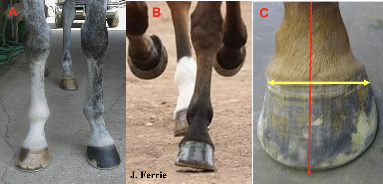
| A Look at Sheared Heels…Often Referred to as ‘Jamming’… Stephen E. O’Grady, DVM 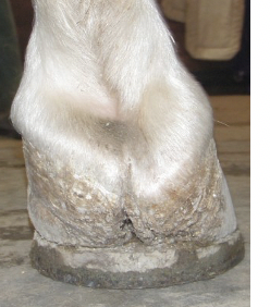 Sheared heels were first described as a problematic foot issue by a veterinarian and a farrier 45 years ago (Moyer and Anderson,1975). There have been many subsequent papers published, many by this author (see here), however, the mechanism of sheared heels is still not completely understood, and the significance of this hoof capsule distortion is underappreciated. Sheared heels are what farriers refer to as ’jamming’. I would often hear farriers talk about ‘jamming’ at the coronet. So, I asked my friend Bill Moyer if he knew what jamming is that farriers’ talk about? He said, "I don’t know about farriery, but jamming is a Latin dance culture practiced in South America." So, then I looked up the definition of jam in the dictionary…1) to become blocked or wedged, 2) to force into a restricted space and 3) to improvise on a musical instrument with a group. As the word jamming doesn’t appear to relate to farriery, perhaps a more professional term such as ‘(proximal) displacement of the coronet’ may be more suitable. Sheared heels were first described as a problematic foot issue by a veterinarian and a farrier 45 years ago (Moyer and Anderson,1975). There have been many subsequent papers published, many by this author (see here), however, the mechanism of sheared heels is still not completely understood, and the significance of this hoof capsule distortion is underappreciated. Sheared heels are what farriers refer to as ’jamming’. I would often hear farriers talk about ‘jamming’ at the coronet. So, I asked my friend Bill Moyer if he knew what jamming is that farriers’ talk about? He said, "I don’t know about farriery, but jamming is a Latin dance culture practiced in South America." So, then I looked up the definition of jam in the dictionary…1) to become blocked or wedged, 2) to force into a restricted space and 3) to improvise on a musical instrument with a group. As the word jamming doesn’t appear to relate to farriery, perhaps a more professional term such as ‘(proximal) displacement of the coronet’ may be more suitable. Over the years, I have spent considerable time repairing quarter cracks which are a common occurrence in competition horses. Interestingly, a quarter crack will generally be associated with a sheared heel. Rather than just repair or patch the defect, I would rather address the cause, hence my interest in pursuing the etiology of this problem. Sheared heels and or quarter cracks do not present a problem in the barefoot horse as the foot appears to adapt; functionally, the hoof capsule at the heels is able to adapt to the uneven footing. However, when shoes are applied this ability to adapt becomes modified. Mechanism A sheared heel is defined as a hoof capsule distortion resulting in a proximal displacement of one quarter /heel bulb relative to the contralateral side of the hoof (Turner 1992). The disparity between the lateral and medial quarter/heel bulb is generally 0.5 cm or more and is measured from the coronet to the ground surface of the foot. When sheared heels was first recognized, it was thought to be caused by excessive trimming on one side of the foot and by limb conformation to a lesser degree. To dispel the first thought, the author did a small study in South Africa. Ten broodmares and ten horses in training were used in the study. To be included, all horses had to have straight carpi and had to land flat on the ground. Horses were trimmed three times at three-week intervals where one side of the foot (usually medial) was excessively trimmed from the toe quarter to the heel. In not one instance could I create the sheared heel foot conformation (O’Grady 1994; unpublished data). It is now generally accepted that sheared heels result from limb conformation and or an offset foot. The most common limb conformation is a horse with a narrow chest with a rotational forelimb deformity (Figure 1A). In motion, the carpus breaks over laterally which causes the horse to land asymmetrically during the landing/stance phase of the stride such that the foot contacts the ground with the lateral side and loads on the medial side (Figure 1B). This asymmetric landing will place a disproportionate load on one side of the foot. The other scenario is the foot that is offset to the lateral side; here the surface area of the ground surface of the foot is decreased when compared to the lateral side (Figure 1C).
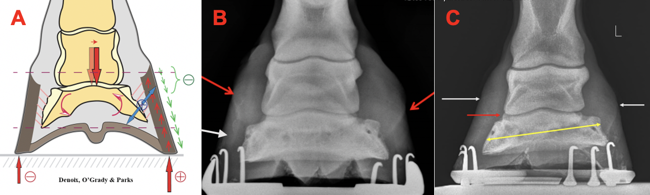
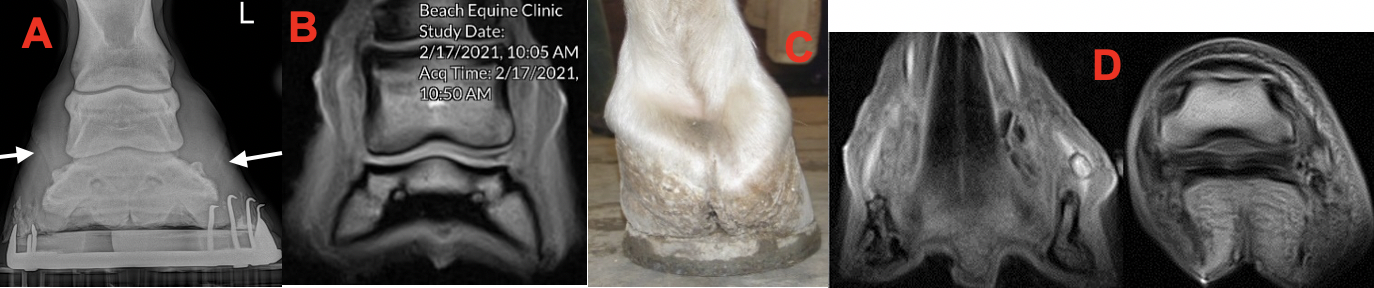
It may be helpful to look at the biomechanical mechanism that occurs on the affected side of the foot. Biomechanically, the position of the coronary band is related to the balance between hoof wall growth at the coronary band and the rate of migration of the hoof wall distally toward the ground. Furthermore, the rate of migration of the hoof wall toward the ground is a balance between an active process occurring in the lamellae to cause them to move distally and the force on the wall from the ground reaction force. Therefore, if the rate of hoof wall growth is greater than the rate of hoof wall migration distally, the coronary band displaces proximally. The increased load on a given side of the foot over time appears to result in biological remodeling rather than the heel being pushed proximally, i.e., the heel is ‘growing’ out of shape rather than being pushed out of shape (Figure 2A). It has been speculated that decreased hoof wall growth or ne growth migrating distally from the coronet may inhibit sole growth, but this has not been proven. The decrease in sole growth may also result from the overload on the affected side. Decreased sole thickness on the affected side will allow the distal phalanx to displace distally…the amount of decent being proportional to the amount of damage to the hoof capsule (Figure 2B & 2C). The change in position of the distal phalanx will generally be noted as a widening of the joint space on radiographs but can also be a compression of the joint space with increased sole depth and or leverage on the contralateral side of the foot (Figure 3A & 3B). Proximal displacement of the heel bulb will also compress the ungual cartilage and soft tissue between the hoof wall and the middle phalanx (Figure 3C & 3D).
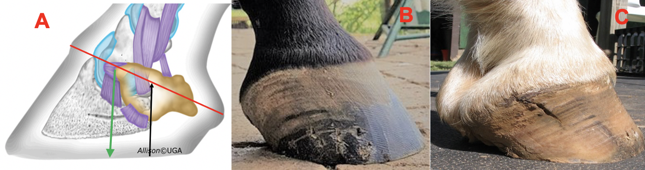
Functionality The heels of the horse's foot have a relatively large amount of flexibility in the proximal to distal (vertical) axis (Figure 4A). This can be explained by the anatomical features of the foot: a discontinuity of the hoof capsule between the heels with highly mobile structures interposed between which are the frog, digital cushion, venous and arterial plexuses, and fibro-cartilaginous connective tissue. In other words, although the dorsal wall is intimately attached to the parietal surface of the distal phalanx, the laminar attachment or suspension in the palmar/plantar section of the foot is far less rigid. This provides the flexibility necessary for function but also allows for proximal displacement of the heels when these structures receive excessive stress or a disproportionate load. We generally see a focal displacement of the coronet that is aligned with the palmar processes of the distal phalanx (Figure 4B & 4C).
Thoughts on farriery While the diagnosis of a sheared heel is straightforward, the etiology of the condition may be misleading and the farriery employed in the treatment is often based on opinions. Sheared heels appear to develop as an adaption-distortion of the hoof capsule because of limb conformation that results in an abnormal strike and loading pattern of the foot on the ground. Farriers often refer to sheared heels as a lateral to medial imbalance or as what I like to refer to as, lateral to medial orientation of the hoof capsule. Many farriers (and veterinarians) feel that a horse should land flat. However, this lateral medial orientation appears to result from the asymmetric landing pattern that is related to limb conformation, therefore, attempting to make the horse land flat would defy conformation and be counterintuitive. Furthermore, a wedge is often applied under the affected side of the foot to make the horse land flat. Considering the biomechanics, elevating one side of the foot would move the CoP toward the side with the wedge and further increase the overload. Interestingly, when the shoes are removed from a foot with sheared heels, the soft tissue structures quickly relax and the conformation changes which gives further evidence of the adaptability of the palmar foot. In fact, when presented with a case of sheared heels, the first part of my farriery, when possible is to remove the shoes for a short period of time (as little as 24 hours is helpful). My farriery goals are to unload the overloaded side of the foot and for this I have used the double trim method for years and have been quite satisfied. Be safe. |