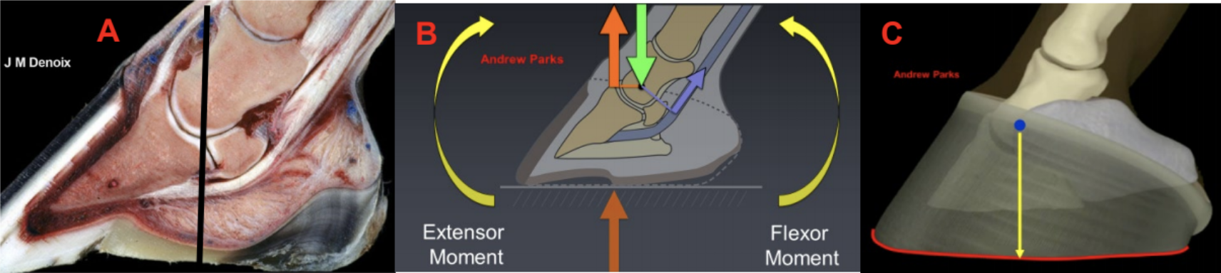
| A Rational Approach to Trimming the Palmar Foot Stephen E. O’Grady, DVM Some of the best criteria for trimming the foot appeared in the literature around the turn of the century. Many of these authors were both veterinarians and farriers…their work based on foot structure and functionality. Recently, there have been many scientific papers published on farriery (often termed evidence-based) but unfortunately, a paucity of information on the appropriate trimming of the equine foot. This author feels that a rational approach to trimming the palmar foot is accomplished by understanding and using the functional anatomy and biomechanics of the foot as guidance and then using good basic farriery principles to implement these guidelines. ‘The horse’s foot is unique as it is a complex of biological structures that follow the laws of physics’ (O’Grady 2009). The equine hoof capsule is comprised of deformable structures…the wall, the sole, the frog and the bulbs of the heels. The structures are continuous with one another, so together they form a coherent, resilient ‘boot’ that encloses the osseous and soft tissue structures of the foot. The functions become obvious as the hoof capsule protects the enclosed structures, allows the foot to accept weight and absorbs concussion and dissipates the energy of impact. To function, the shape of the hoof capsule is a truncated cone. The horn tubules grow dorsally at anywhere between a 45–60-degree angle at the toe and the angle at the heel, generally lower than the toe, dictated by the conformation and integrity of the soft tissue structures in the palmar/plantar foot. The direction of growth of horn tubules means the heels can’t grow tall…they must grow forward. Therefore, in farriery, the term ‘lower the heels’ may be a misnomer and should be replaced with ‘trimming the heels’ which creates ground surface. If we consider the anatomy, the foot is divided into dorsal and palmar/plantar sections…the rigid dorsal section is designed to accept the weight of the horse and the palmar/plantar section to absorb the impact with the ground (a generalized statement as there is a myriad of interactions that occur within the soft tissues during weight bearing) (Figure 1A). To insure proper physiological function of the foot, both sections interact with each other, complement each other, and must be included within the hoof capsule to work together. If the ground surface of the palmar section of the hoof capsule is decreased, the soft tissue structures in the palmar/plantar foot are no longer included within the hoof capsule. Therefore, these structures are bypassed during the landing phase of the stride and the load is placed on the lamellae and bones of the digit. Furthermore, when the soft tissue structures are enclosed within the hoof capsule, weight bearing is distributed over the entire foot. The soft tissue structures do not have the ability to accept full weight but can share weight with the heels of the hoof capsule. The equine foot rotates around a fixed point which is the center of rotation COR. Weight is borne through the COR and is opposed by an equal and opposite force (GRF). However, these two forces are not aligned, and this creates a moment about the distal interphalangeal joint (the extensor moment). For the foot to be stable on the ground at rest, this moment will have an opposing moment created by the deep digital flexor tendon (the flexor moment) (Figure 1B). For an overview of biomechanics, please see here. When heels are allowed to migrate dorsally, the weight placed on the palmar/plantar foot is now borne on a decreased surface area. Depending on the structures of the palmar foot, this overload may lead to collapse of the heels which in turn increases the tension in the DDFT (flexor moment). The increase in the flexor moment results in the GRF moving dorsally leading to an increase in the extensor moment. Finally, using the knowledge of anatomy and an understanding of biomechanics, farriers can trim appropriately utilizing the COR to promote/maintain a healthy functional hoof capsule (Figure 1C).
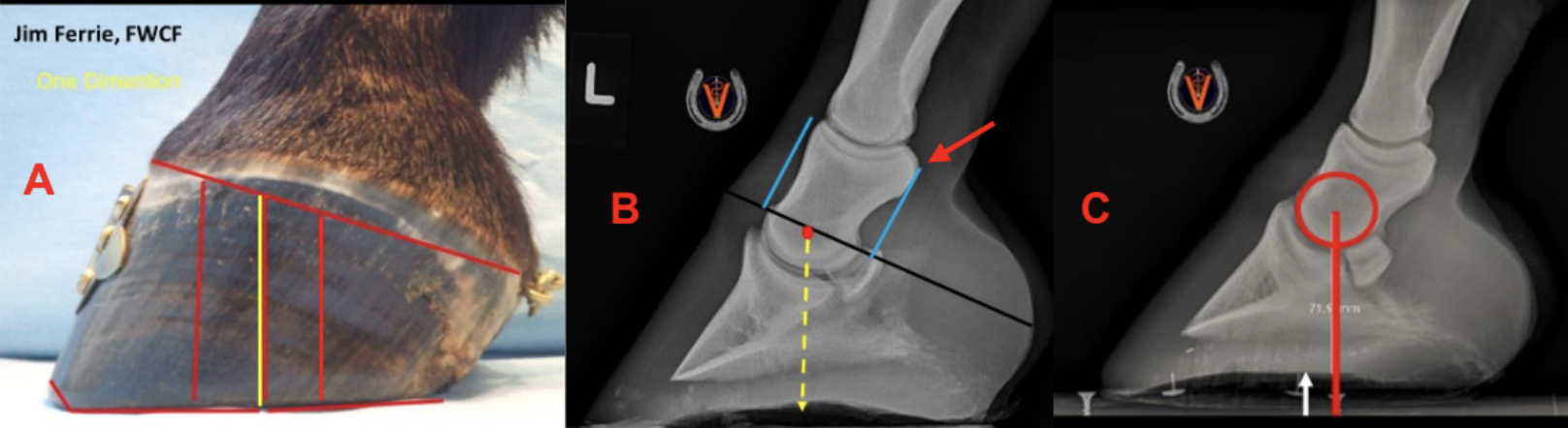
The Trim The author has always used the hoof-pastern axis, the center of rotation (COR) and the heels trimmed to the base of the frog or to the same plane as the frog as guidelines for the trim. Please see here. For our discussion here, I will only consider the center of rotation. Recently, there have been multiple ‘mapping’ protocols described to act as guidelines for trimming, all correlating with the COR; however, I have not found them to be consistently repeatable and somewhat cumbersome to use on a routine basis. One method that I have found repeatable is one described by farrier Jim Ferrie, FWCF (Figure 2A). Here, the coronary band on the lateral side of the hoof capsule is divided into thirds. The dorsal and palmar/plantar border of the middle phalanx (P2) can be palpated easily in the standing horse. The tuberosity at the dorsal palmar border can be palpated and used as a landmark. If a line parallel to the borders of the bone is drawn to the coronet and then a vertical line is drawn from the center of these two lines at the coronet to the ground, this will approximate the COR (Figure 2B). Recently, the author measured the widest part of the foot in a group of horses with varying foot conformation. A line was drawn across the foot at the widest part, a radiopaque marker was placed, and a lateral radiograph was taken…making sure that MC3 was perpendicular to the ground. It was discovered that in all instances, the widest part of the foot was 6mm-9mm dorsal to the COR (Figure 2C) (O’Grady and Allen 2016 unpublished data).
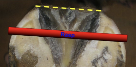 Appropriate or consistent trimming of the palmar/plantar section of the foot has always presented problems for the clinician, for a variety of reasons. Variations in hoof conformation, farrier training, empiric considerations, and owner pressure not to trim the heels (low) often dictate how the heels are trimmed. If the heels are not trimmed appropriately and can migrate dorsally toward the center of the foot, the laminar interface and the bones of the digit assume the function of the soft tissue structures. Often, the heels of the hoof capsule can’t be trimmed to the base of the frog for anatomical reasons or due to structural damage, but an attempt should always be made to trim the heels of the hoof capsule to the same horizontal plane as the frog. If the hoof wall at the heels cannot be trimmed to the base of the frog, the branch of the shoe or some other form of farriery can be used to extend the ground surface of the hoof capsule to the base (or slightly beyond) of the frog. If the frog lies distal to the ground surface of the foot, increased load is placed on the soft tissue. In the opposite scenario, if the frog is recessed below the surface of the foot, increased load is placed on the heels of the hoof capsule. In either case, the author is going to initiate the appropriate farriery to return the frog to the same plane as the heels of the hoof capsule which then makes the palmar/ plantar section of the foot ‘load sharing’. In doing so, ‘farriery becomes not only an art but truly a science’. Appropriate or consistent trimming of the palmar/plantar section of the foot has always presented problems for the clinician, for a variety of reasons. Variations in hoof conformation, farrier training, empiric considerations, and owner pressure not to trim the heels (low) often dictate how the heels are trimmed. If the heels are not trimmed appropriately and can migrate dorsally toward the center of the foot, the laminar interface and the bones of the digit assume the function of the soft tissue structures. Often, the heels of the hoof capsule can’t be trimmed to the base of the frog for anatomical reasons or due to structural damage, but an attempt should always be made to trim the heels of the hoof capsule to the same horizontal plane as the frog. If the hoof wall at the heels cannot be trimmed to the base of the frog, the branch of the shoe or some other form of farriery can be used to extend the ground surface of the hoof capsule to the base (or slightly beyond) of the frog. If the frog lies distal to the ground surface of the foot, increased load is placed on the soft tissue. In the opposite scenario, if the frog is recessed below the surface of the foot, increased load is placed on the heels of the hoof capsule. In either case, the author is going to initiate the appropriate farriery to return the frog to the same plane as the heels of the hoof capsule which then makes the palmar/ plantar section of the foot ‘load sharing’. In doing so, ‘farriery becomes not only an art but truly a science’. Be Safe… |