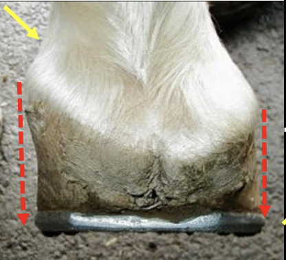
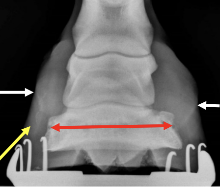
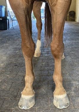
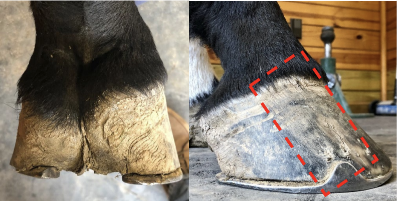
| Sheared Heels: A Case Report Stephen E. O’Grady, DVM Sheared heel conformation is a hoof capsule distortion fraught with controversy. It was first described by a veterinarian and a farrier forty-five years ago and still being debated (Moyer and Anderson 1975). A sheared heel is defined as a hoof capsule distortion resulting in a proximal displacement of one quarter/heel bulb relative to the contralateral side of the hoof (Turner 1992) (Figure 1).The distortion is continually characterized as a lateromedial hoof imbalance, however; the author feels it should be considered an adaptation to unequal forces caused by limb conformation which promotes an asymmetric landing pattern when the foot strikes the ground (O’Grady and Castelijns 2011). Furthermore, the hoof capsule distortion can be exacerbated by various farriery attempts to create a flat landing pattern which opposes the limb conformation. For an in-depth look at sheared heels, please follow this link. Briefly, the equine foot can change shape or adapt when disproportionate forces are placed on the foot during landing. This adaptation can be seen radiographically where one heel bulb is displaced proximally relative to the other side while the distal phalanx remains parallel with the ground (Figure 2) Sheared heels are generally seen with horses that have a rotational deformity involving the forelimbs or when the foot is markedly offset toward the lateral side. With a rotational deformity, the carpus will breakover in a lateral direction which creates an arc as the foot approaches the ground for weight bearing (Figure 3). The foot will then strike the ground asymmetrically, overloading one heel bulb and displacing it proximally. When a sheared heel becomes excessive, lameness can occur due to bruising, hoof wall separations, quarter cracks and soft tissue trauma dorsal to the coronet. Appropriate farriery will address the hoof capsule distortion and will unload the section of the foot that is being displaced. Attempting to make the foot land flat with trimming and shoeing should be discouraged as it defies the horse’s limb conformation and makes the problem worse.
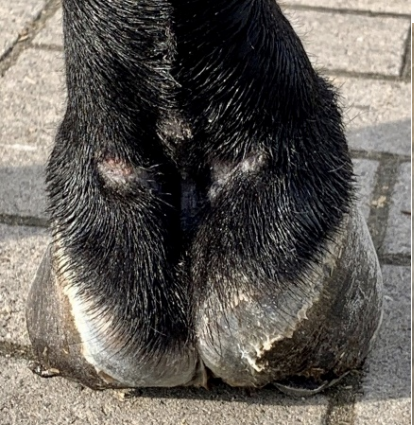
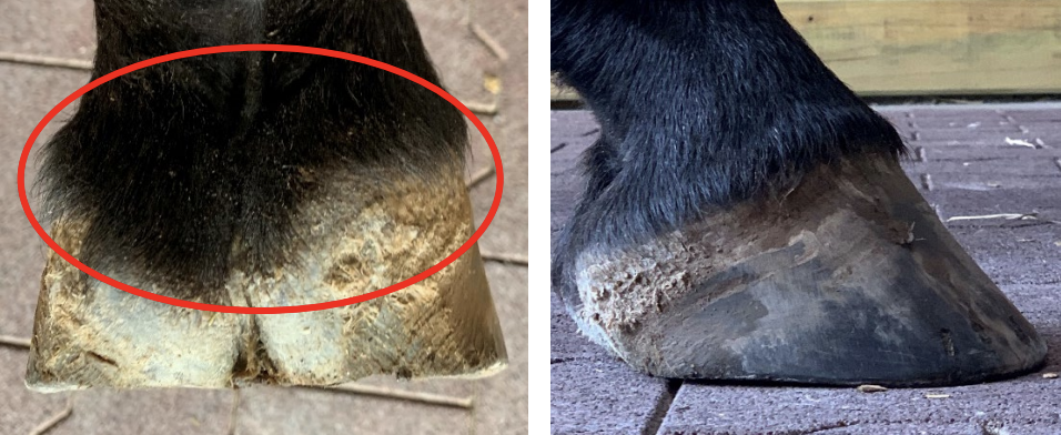
Case Report A 9-year-old gelding was imported from Europe in December 2019. On arrival, he had a marked sheared heel on the lateral side of the RF foot (Figure 4). The horse was subsequently sent to Florida for the Winter Festival where he showed successively as a jumper. During his time in FL, the forefeet showed some improvement with the trainer’s farrier, however; when the horse returned to Virginia, the trainer enquired whether the foot conformation could be improved further (Figure 5).
I have always been a big proponent of rehabilitating the palmar foot in a barefoot state in the initial stages when possible. For more information on barefoot methodology, please follow this link. Removing the shoes will reposition the frog, place the heels in a uniform ‘weight sharing’ situation, relieve the compression of the soft tissue above the coronet on the displaced heel and allow the structures of the palmar foot to assume a more acceptable conformation. With the COVID 19 pandemic and no horse shows on the horizon, it was agreed to give the horse a month off. The shoes were pulled, the solar surface of the foot was cleaned briskly with a wire brush, the heels of the hoof capsule were trimmed to the same plane as the frog and the perimeter of the hoof wall was rounded using a bevel method. The horse was walked daily on a firm surface and then turned out in a small paddock. At no time are boots ever used as ‘boots are not barefoot’ and will hinder the rehab process. At 2 weeks, the farrier returned and again trimmed the heels from the widest part of the foot to assure they were on the same plane as the frog (Figure 6).
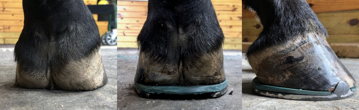
Four weeks later the horse was shod using the double trim method. The heels were again trimmed to the same plane as the frog, toe length was reduced, and any flares were removed from the outer hoof wall. A steel shoe was fitted with a stiff pad to stabilize and maintain the conformation of the heels. A second trim was performed on the displaced side in a tapered fashion from the toe quarter to the heel. This creates a space between the hoof and the shoe (pad) to further unload the heel and let it drop further. When attaching the shoe, only one or perhaps two nails are used in the toe of the shoe, so the displaced heel is not prevented from dropping down to the shoe (Figure 7).
Assessing the limb conformation, observing the foot landing, and improving the foot shape utilizing the appropriate farriery appear to be the important aspects of addressing sheared heels. Some Thoughts The palmar section of the horse's foot has a relatively large amount of flexibility in the proximal to distal (vertical) axis. This can be explained by the anatomical features of the foot: a discontinuity of the hoof capsule between the heels which are interposed with highly mobile structures which are the frog, digital cushion, ungual cartilages, venous and arterial plexuses and fibro-cartilaginous connective tissue. Stated another way, although the dorsal wall is intimately attached to the parietal surface of the distal phalanx by the lamellae, the laminar attachment or suspension in the palmar/plantar section of the foot is far less rigid. This provides the flexibility necessary for function but also allows for proximal displacement of the heels when these structures receive excessive stress or a disproportionate load. Functionally, this arrangement serves the bare-footed horse well as the hoof capsule at the heels can adapt to the uneven footing, however; when shoes are applied this ability to adapt becomes modified. Therefore, when trimming or shoeing modifications in the sagittal plane of the foot are being contemplated, it is important to be aware of this vertical mobility, and the tendency for vertical displacement of the heels. I would like to recognize farrier Jose Lopez for his excellent farriery on this case. |