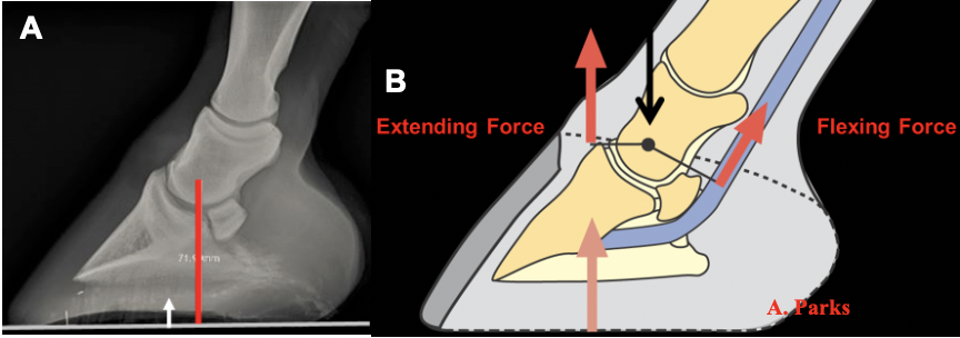
| Using Radiographs as a Guideline for Farriery Stephen E. O’Grady, DVM Using foot radiographs to apply the appropriate farriery is an ideal way of ‘forging’ a relationship between the veterinarian and farrier. When the two professions work together to interpret and apply the information garnered from foot radiographs; it really forms a ‘symbiotic’ relationship. Tasked with applying the appropriate farriery, the farrier can be assisted by “radiographically” seeing within the hoof capsule. This is invaluable for determining the best possible trim as well as the precise design & placement of any proposed shoe or appliance. Therefore, it becomes important for the veterinarian and farrier to acquire the necessary knowledge and expertise to use foot radiographs as a template or guideline to apply the appropriate farriery. To transfer the information from a foot radiograph to the foot, one must have a consistent starting point or reliable landmark. This author has always used the widest part of the foot (or some may prefer the widest part at sole-wall junction) as my starting point. It must be remembered that the position of the distal phalanx remains constant while the deformable hoof capsule changes shape. To give the widest part of the foot more credibility, we did a small impromptu study where the widest part of the foot was measured and marked on several horses with varying foot conformation and then radiographed. In all cases the widest part of the foot was located 5-10 mm dorsal to the center of rotation (COR) (Parks 2011; O’Grady and Allen 2017) (Figure 1A). Therefore, the widest part of the foot appears to be, not only an appropriate landmark but utilizes the COR to apply the relevant biomechanics to the foot (Figure 1B).
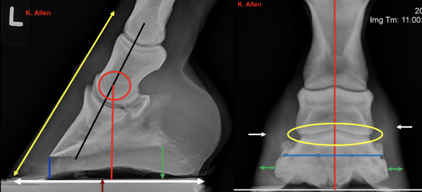
Foot radiographs in the veterinary literature is termed a podiatry study. The podiatry study consists of a lateral to medial view (lateral) and a dorso palmar 0° view (DP). Other than any pathology noted on either view, the following guidance for farriery should be noted: the lateral view shows axial alignment, the hoof-pastern axis, the A B A. Parks center of rotation, sole depth / thickness, the angle of the solar border of the distal phalanx and the proportions of the ground surface on either side of the COR. The DP view shows the alignment of the digit, the orientation of the distal phalanx in the frontal plane, disparity in the joint spaces, thickness of the hoof wall as well as the position of the coronet on either side of the distal phalanx (Figure 2). It should be noted here that the preparation of the foot, the proper positioning of the foot and the appropriate radiographic technique when taking the radiographs is of the utmost importance to assure quality images. Virginia Equine Imaging has discussed “How To’ acquire consistent quality radiographic images. Please see…
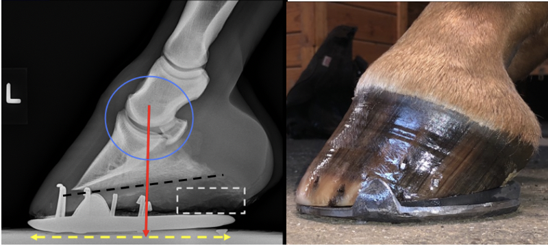
Let us use a couple of random examples to show how we can use radiographs as a guide to apply farriery. More examples will follow… Example 1: This radiograph shows a Grade 2 (2-4) clubfoot. The horse had a marked shortened cranial phase of the stride on that limb. The distal interphalangeal joint (DIPJ) shows a mild flexural deformity which places increased load on the dorsal rim of the DIPJ, there is adequate sole depth but an increase in palmar angle and a shoe that is too small. The radiolucent area in the palmar section of the foot indicates a recessed frog placing excessive load on the hoof wall. To load the heels and place the hoof wall and frog on the same plane, the heels should be trimmed from the middle of the foot palmarly. Following the trim, with the increased ground surface, the foot will require a larger shoe and possibly heel elevation to address any stress placed on the DDFT by the trim. A picture following the proposed farriery is included below.
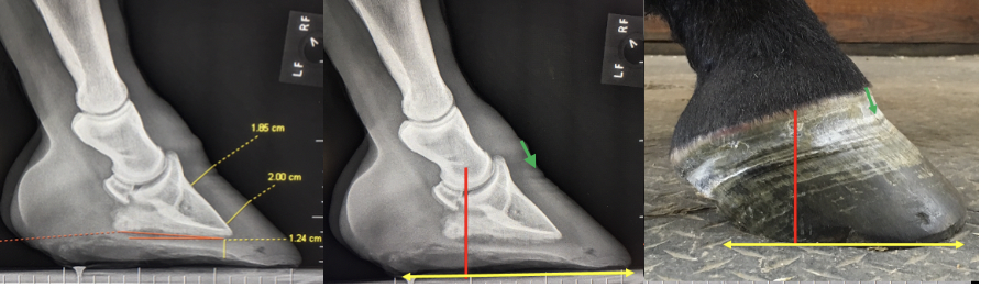
Example 2: This radiograph was sent to my consulting service on a lameness case. The measurements on the radiograph are helpful to show distortions, comparing studies, record keeping and perhaps client information. However, I find them difficult to use as a guide to apply farriery especially when the difference in numerical values are minute. The image shows a long toe (flare) along with the heels migrating dorsally forming a ‘knob’ shape to the heel bulbs. The coronet at the toe shows the direction of hoof wall growth distally, packed growth rings (a sign of stress) and minimal disparity in hoof wall growth from toe to heel. The second image shows guidelines inserted for the farrier. Note that there is adequate sole depth present so any reduction in toe length should only be in a vertical direction or from the outer hoof wall. The third picture shows the guidelines applied to the foot and the issues noted on the radiograph can be appreciated on the foot.
For further information on foot radiographs, please see here. Be / Stay safe! |