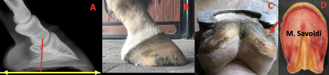
| Farriery For The Hind Feet – An Ongoing Conundrum Stephen E. O’Grady, DVM The hind feet in the horse have not been studied in comparison to the forefeet. The function of the forefeet is weight bearing which is associated with foot lameness while the function of the hindfeet is propulsion. It may be a misnomer to think that the force generated by the hind feet during propulsion does not exert an effect on the soft tissue structures / joints on the upper body and back of the horse. Recent studies have shown a correlation between abnormal hoof conformation in the hind feet and hind limb lameness localized above the foot (Clements PE et al 2020; O’Grady et al 2018; Pessanite L et al 2018). However, it must be remembered that a correlation does not equal causation. Causation is a predictable action that can cause another action or problem while correlation is an action, although not certain, may have a direct link to another action or problem. Abnormal Hindfoot Conformation 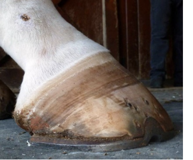 The low heel “bull nose” conformation of the hind foot has become so prevalent in farriery that it is often considered normal (see insert). This abnormal conformation of the hind feet is easy to recognize. When looking at the limb from the side, the digit will show a broken back hoof-pastern axis. The slope of the coronary band from the toe to the heel will have an acute angle of 40–45°, and the coronet will bend distally at the heel to become almost vertical. The bulbs of the heels will be prolapsed plantar to the heels of the hoof capsule and will form a “knob” shaped appearance that can be seen lying against the shoe. The hair on the coronet at the heels will often project horizontally rather than lying flat against the hoof due to the excessive load on the associated hoof wall. There will be a disparity in the growth rings below the coronet from the toe to the heel, with the growth rings wide apart at the toe and then tightly packed at the heel. Remembering that packed growth rings are generally a sign of overload when evaluating distortions. The dorsal hoof wall will assume a “bull nose” appearance. Looking at the foot from behind, the frog will be large and bulbous from the constant stimulation with the ground, a ledge will form in the frog from bearing weight, and it will be situated well below the hoof wall with the bulk of the frog now located between the two branches of the shoe if shod. The solar surface of the foot will show an inclined plane of the entire frog from the base to the apex in a dorsal cranial direction toward the coronet. This inclined plane or angle will match the angle of the solar border of the distal phalanx in the hoof capsule. The toe area on the solar surface of the foot will show a deep or exaggerated concavity between the apex of the frog and the inner branch of the shoe instead of a steep yet smooth transition of the sole from the frog to the sole wall junction. There will usually be a palpable “trough” located just dorsal to the apex of the frog. Upon removing the shoe, the end of the heel of the hoof capsule is located well forward from the base of the frog and the horn tubules will be parallel with the ground. The hoof wall at the heel will be thin, the bars may be damaged or missing, and the angle of the sole will be absent. Lightly paring the area adjacent to the hoof wall at the end of the heel with a hoof knife will often show moderate to severe hemorrhage from the pressure of the damaged hoof capsule against the shoe. When the foot is placed on the ground, total weight bearing will be placed on the frog, which is located distal to the ground surface of the hoof capsule, and many horses will be reluctant to stand on it when the opposing limb is lifted off the ground. As noted previously, hoof testers placed on either side of the heel at the angle of the sole will often elicit a painful response and the structures will deform (Figure 1). The low heel “bull nose” conformation of the hind foot has become so prevalent in farriery that it is often considered normal (see insert). This abnormal conformation of the hind feet is easy to recognize. When looking at the limb from the side, the digit will show a broken back hoof-pastern axis. The slope of the coronary band from the toe to the heel will have an acute angle of 40–45°, and the coronet will bend distally at the heel to become almost vertical. The bulbs of the heels will be prolapsed plantar to the heels of the hoof capsule and will form a “knob” shaped appearance that can be seen lying against the shoe. The hair on the coronet at the heels will often project horizontally rather than lying flat against the hoof due to the excessive load on the associated hoof wall. There will be a disparity in the growth rings below the coronet from the toe to the heel, with the growth rings wide apart at the toe and then tightly packed at the heel. Remembering that packed growth rings are generally a sign of overload when evaluating distortions. The dorsal hoof wall will assume a “bull nose” appearance. Looking at the foot from behind, the frog will be large and bulbous from the constant stimulation with the ground, a ledge will form in the frog from bearing weight, and it will be situated well below the hoof wall with the bulk of the frog now located between the two branches of the shoe if shod. The solar surface of the foot will show an inclined plane of the entire frog from the base to the apex in a dorsal cranial direction toward the coronet. This inclined plane or angle will match the angle of the solar border of the distal phalanx in the hoof capsule. The toe area on the solar surface of the foot will show a deep or exaggerated concavity between the apex of the frog and the inner branch of the shoe instead of a steep yet smooth transition of the sole from the frog to the sole wall junction. There will usually be a palpable “trough” located just dorsal to the apex of the frog. Upon removing the shoe, the end of the heel of the hoof capsule is located well forward from the base of the frog and the horn tubules will be parallel with the ground. The hoof wall at the heel will be thin, the bars may be damaged or missing, and the angle of the sole will be absent. Lightly paring the area adjacent to the hoof wall at the end of the heel with a hoof knife will often show moderate to severe hemorrhage from the pressure of the damaged hoof capsule against the shoe. When the foot is placed on the ground, total weight bearing will be placed on the frog, which is located distal to the ground surface of the hoof capsule, and many horses will be reluctant to stand on it when the opposing limb is lifted off the ground. As noted previously, hoof testers placed on either side of the heel at the angle of the sole will often elicit a painful response and the structures will deform (Figure 1).
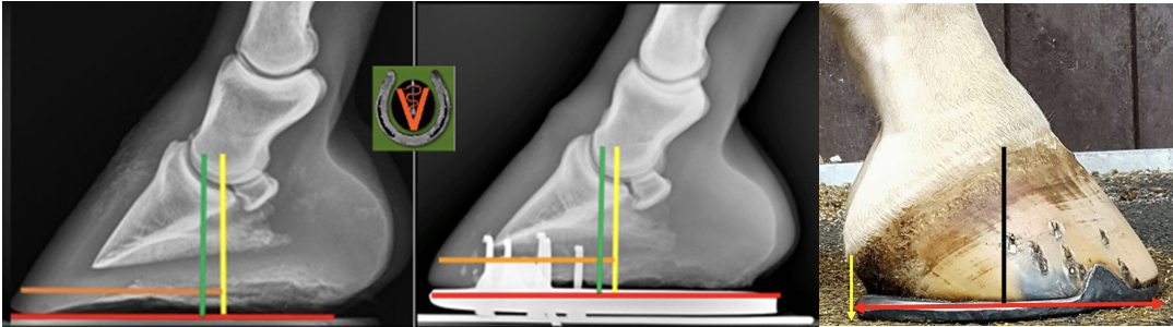
Etiology I don’t recall this abnormal hindfoot conformation to be an issue when I was a practicing farrier or during my early veterinary career. It is always easier to understand a phenomenon if one can establish a cause. The biomechanics of the hindfoot has not been evaluated when compared to the front foot. I did a small impromptu study using hind foot radiographs from two large equine practices for a recent paper and found some significant differences when compared to the fore foot. Using lateral radiographs summitted by two equine radiologists that were considered to be good hind foot conformation as a model; it was noted that the center of rotation (COR) was located further plantar than the forefoot and the hoof-pastern axis was mildly broken back (Figure 2).
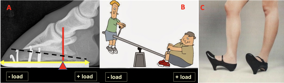
With the anatomy in mind, it may be possible to deduce a possible etiology. If the heels of the hoof capsule are not trimmed appropriately and left to migrate dorsally, it will further decrease the ground surface of the foot plantar to the COR. As the plantar ground surface diminishes, the frog will prolapse, descend distally below the ground surface of the foot, and then becomes large / bulbous. The load is now being placed on the soft tissue structures and the plantar section of the foot becomes overloaded. The decreased ground surface in the plantar foot coupled with a broken back hoof-pastern axis causes a disproportionate load on the bottom of the foot. This is further evidenced by the increased sole thickness in the dorsal foot and the margin of the distal phalanx migrating toward the dorsal hoof wall creating the ‘bull nosed’ conformation. As the ground surface of the plantar foot diminishes and the hoof capsule distortion worsens, the limb is moved dorsally under the horse which can often be viewed as a mild ‘sickle’ hock conformation. The abnormal hoof conformation coupled with the position of the limb, creates excessive stress on the soft tissue structures and joints of the hind limb (Figure 3).
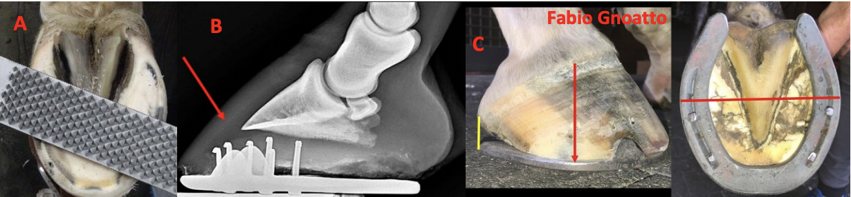
Thoughts on Farriery The hind feet seem to improve / restore better when compared to the forefeet. One reason is that it appears the frog and digital cushion are displaced in the hind foot rather than damaged from weight bearing in the forefoot. The first step in rehabilitating the hind foot is to get the plantar structures ‘load sharing’ again i.e., getting the heels of the hoof capsule and the frog on the same plane. The easiest way I have found is to remove the shoes and allow the horse to be barefoot for a brief period (even for a few days). The next step is to trim the toe and remove the cavitation in the dorsal sole. Even though there marked sole thickness noted on the radiograph, the farrier will often encounter some hemorrhage when trimming the hoof wall at the toe. The reason for this is that as the distal phalanx migrates toward the dorsal hoof wall, it stretches the dermis which is often misleading on a radiograph. Finally, when fitting the shoe, draw a line across the widest part of the foot and fit the shoe such that the line is in the middle of A B C the shoe and there are approximate ground surface proportions on either side of the line. The branches of all my hind shoes extend to a point where a vertical line drawn from the end of the reaches the hair line at the heel bulb (Figure 4).
For further reading on farriery for the hind foot, please see here. Be Safe |